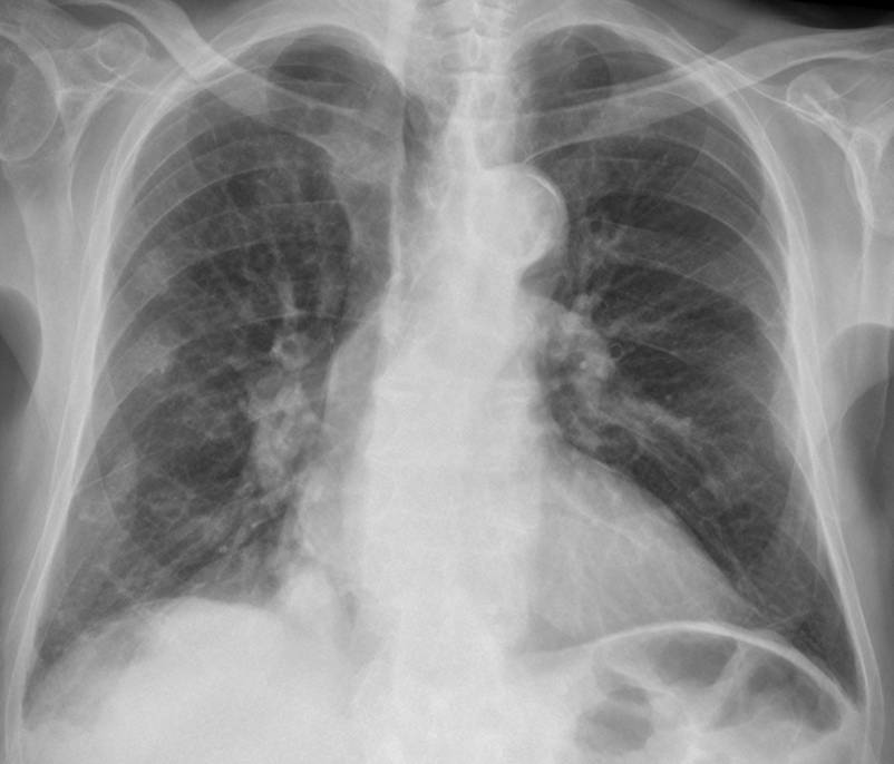
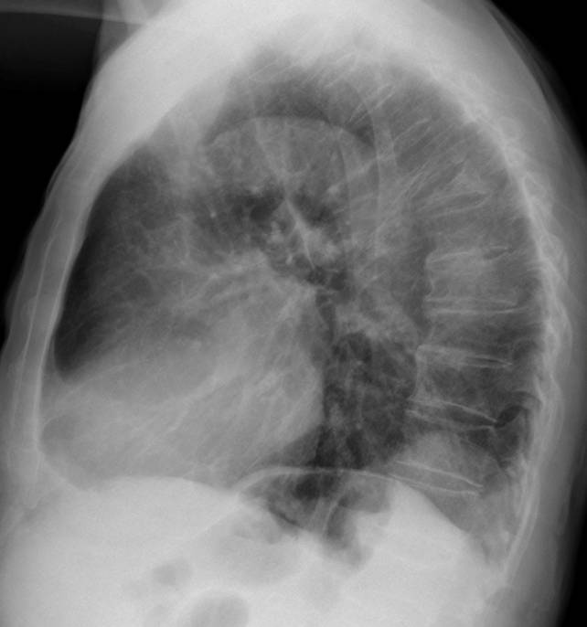
Showing chest radiographs of an 81-year-old male with multiple bone fractures after a car accident. There is a rounded well-defined opacity in the posterior costophrenic sulcus. What do think it is?
1. Carcinoma of the lung
2. Bochladek’s hernia
3. Diaphragmatic cyst
4. All of the above
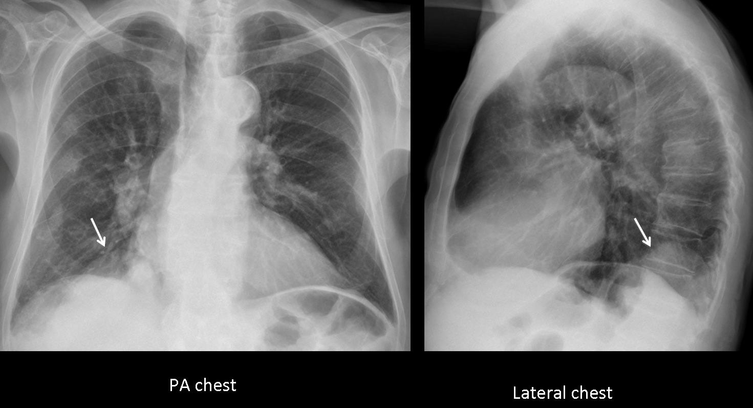
Fig. 1
Findings: aside from multiple rib fractures and a T-6 compression fracture, there is a bulge in the posterior aspect of the right hemidiaphragm (arrows).
The diaphragm is a frontier organ, and localised bulges in its contour may be due to alterations of the diaphragm itself (eventration), pleural or lung tumours, or a condition arising from below (hernia).
The most common lesion in this area is Bochdalek hernia, which is not infrequent in elderly people. However, a diaphragmatic cyst cannot be ruled out because conventional chest radiography cannot distinguish among the densities of fat (hernia), fluid (cyst), and soft tissue (tumour). A well-defined lung tumour cannot be excluded because the costophrenic space is narrow and the differential characteristics of intra- vs. extra-pulmonary lesions cannot be applied.
Therefore, the correct answer is: 4. All of the above
In this particular case, coronal and sagittal CT demonstrate a necrotic pulmonary mass occupying the costophrenic angle (arrows). Diagnosis: carcinoma of the lung, confirmed at surgery.
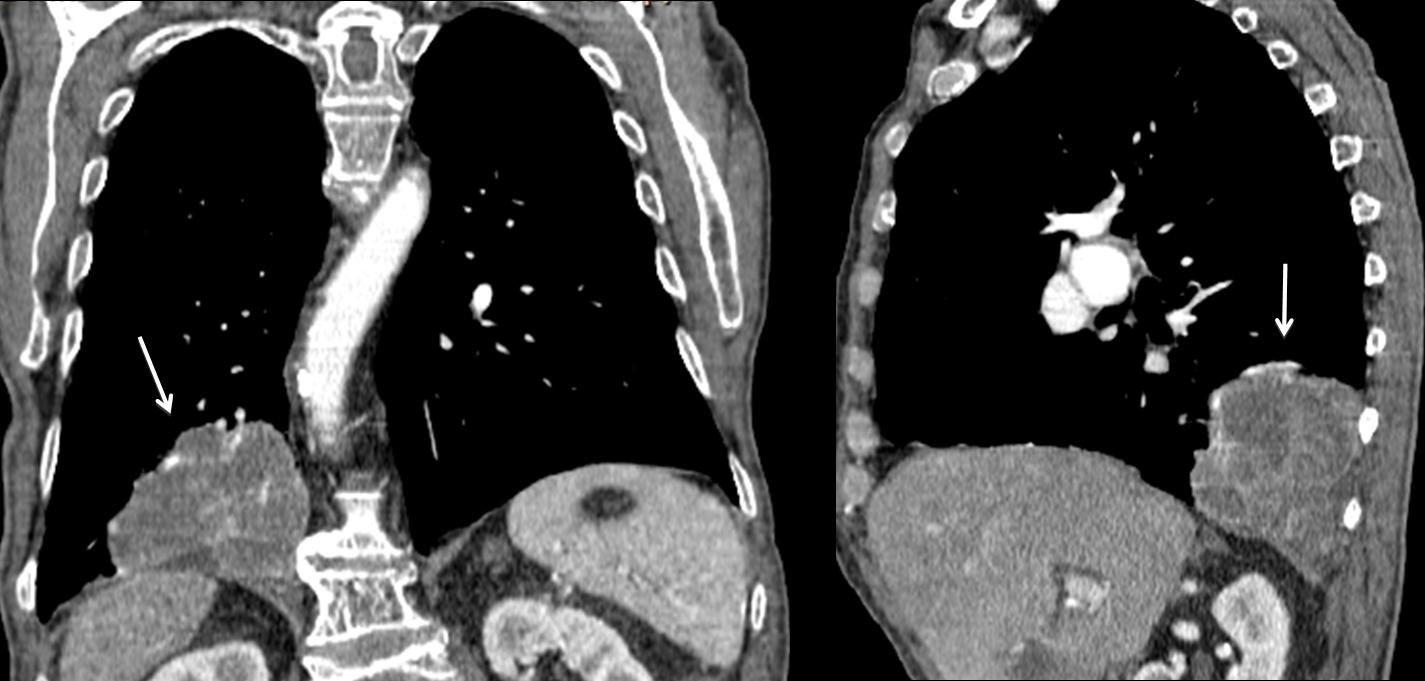
Fig. 2
Occasionally, lung tumours may develop in the posterior costophrenic sulcus, as in the case below, which corresponds to an asymptomatic 56-year-old man. Chest radiographs show findings similar to the previous case, with a rounded opacity in the posterior sulcus (Fig. 3, arrows) which simulates diaphragmatic pathology.
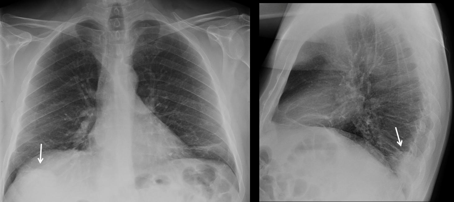
Fig. 3
Coronal and sagittal CT demonstrate a rounded lung tumour (Fig. 4, arrows), separated from the diaphragm.
Final diagnosis: carcinoma of the lung
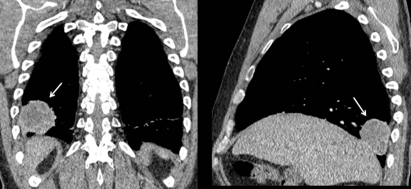
Fig. 4
By far, the most common lesion causing a bulge in the posterior costophrenic sulcus is Bochdalek hernia. Acquired Bochdalek hernia is due to weakening of the diaphragmatic muscle fibres, with protrusion of abdominal fat. The incidence of this condition increases with age and it is not an uncommon finding in asymptomatic elderly individuals.
In Fig. 5, Bochdalek hernia appears as a rounded posterior sulcus lesion (arrows). Note imaging similarity to previous cases.
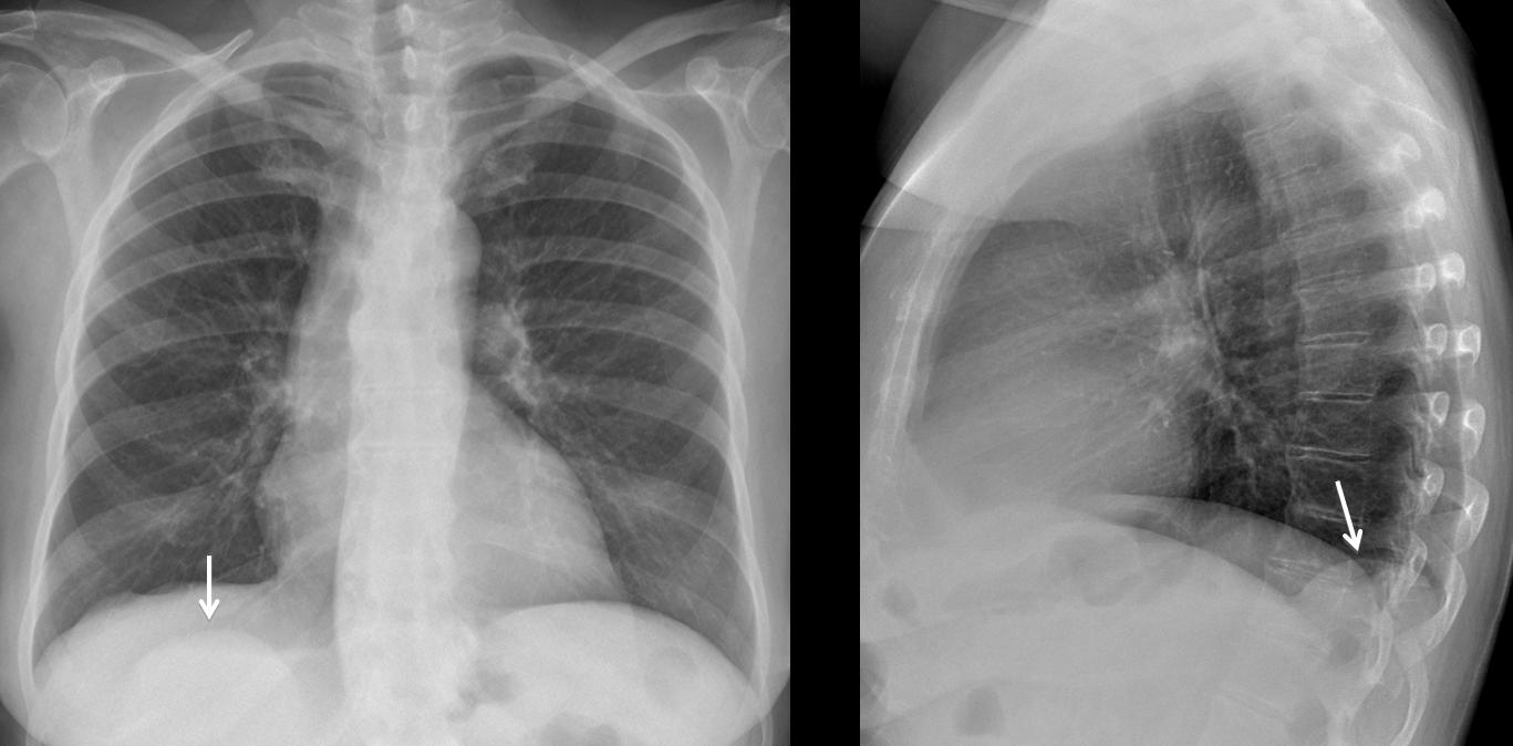
Fig. 5
The diagnosis is confirmed by axial CT, which shows the herniated fat (arrow). Sagittal CT confirms fat herniation through a diaphragmatic hiatus (arrow).
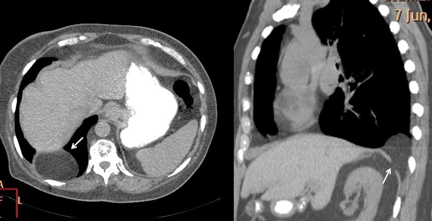
Fig. 6
Bilateral Bochdalek hernia is not uncommon. In this 72-year-old man, chest radiographs show posterior masses in both costophrenic angles (Fig. 7, arrows)
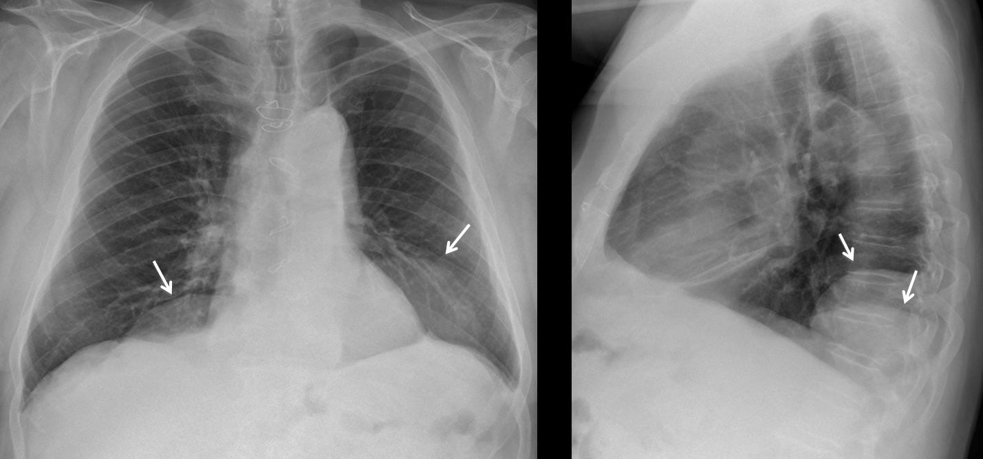
Fig. 7
Coronal and sagittal CT confirm the bilateral herniated abdominal fat (arrows) and posterior discontinuity of the diaphragm (red arrow).
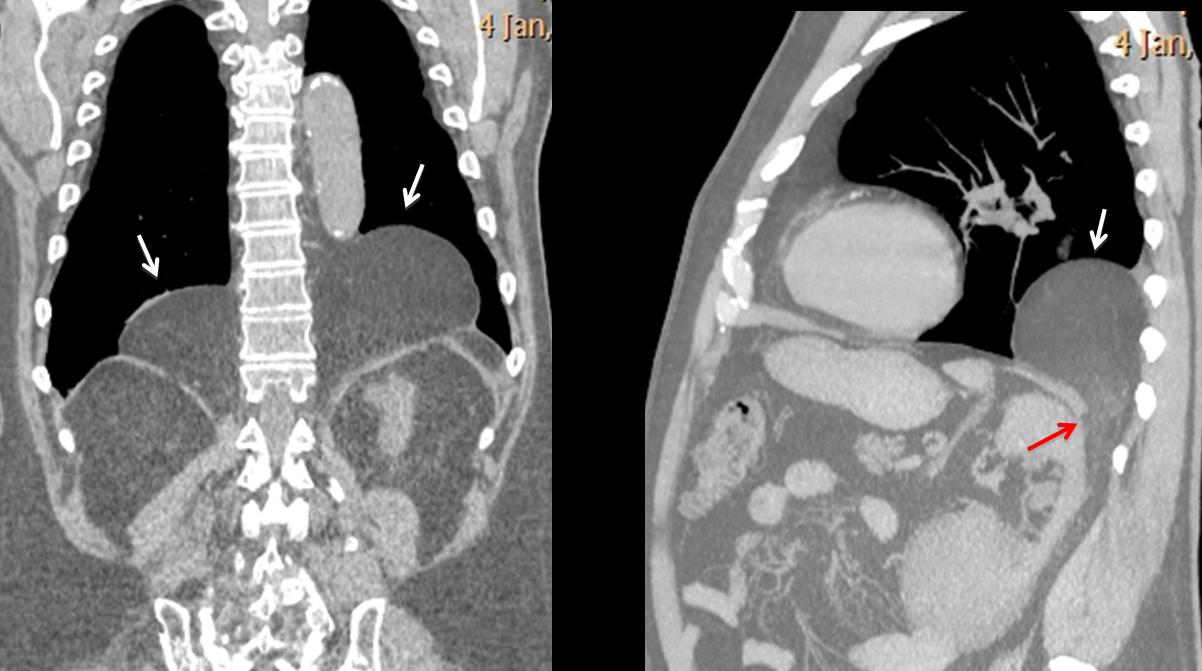
Fig. 8
Diaphragmatic masses are uncommon and cannot be distinguished from other lesions in chest radiographs. A solid mass attached to the diaphragm is likely to be a localised fibrous tumour of pleura. Diaphragmatic cysts are very rare and can be diagnosed by CT. Fig. 9 shows a diaphragmatic bulge (arrows) in a 61-year-old woman that has remained unchanged for the last ten years.
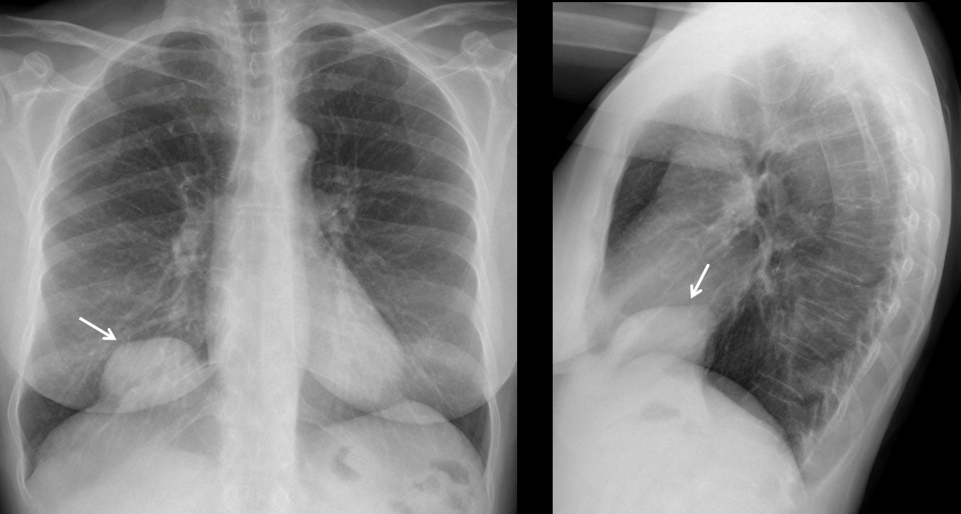
Fig. 9
On unenhanced CT, the mass shows fluid content and peripheral calcifications.
Final diagnosis: diaphragmatic cyst.
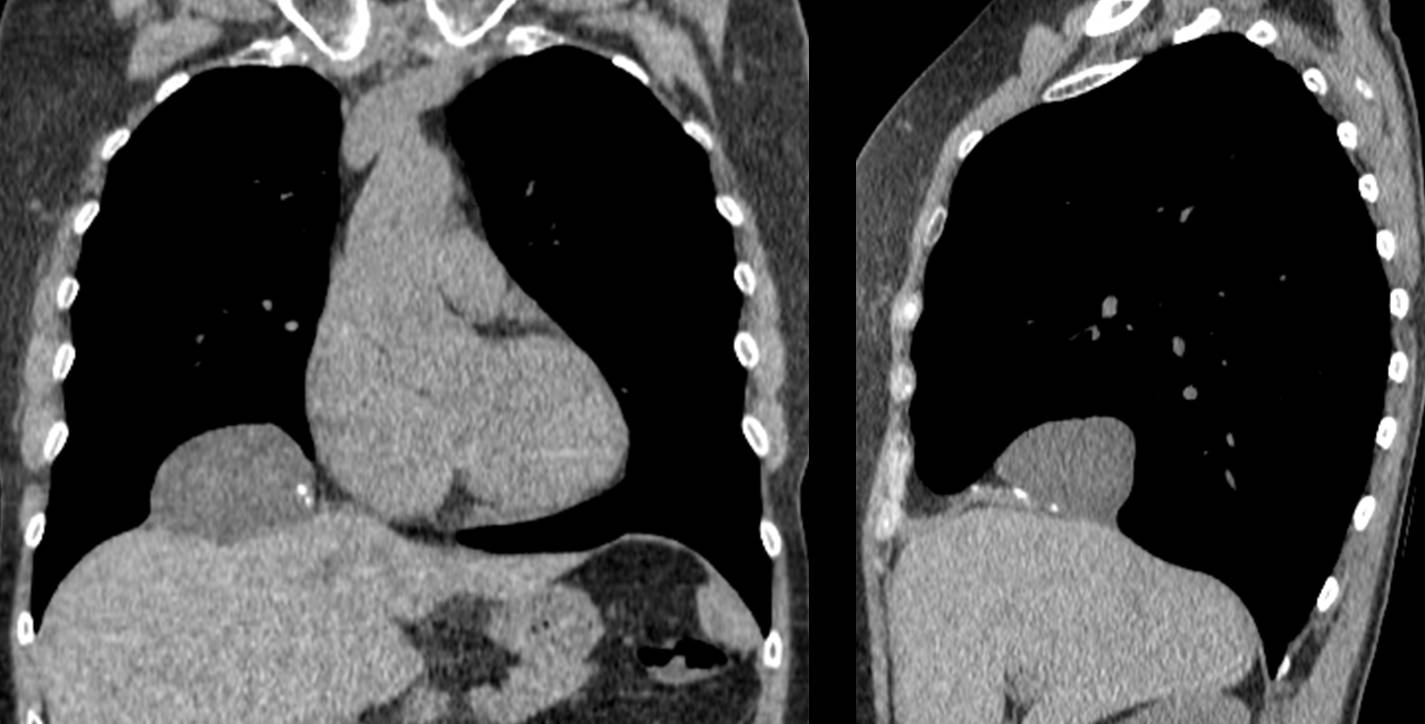
Fig. 10

Follow Dr. Pepe’s advice
Follow Dr. Pepe’s advice:
- Diaphragmatic bulges may be related to a diaphragmatic condition, or reflect disease above or below the diaphragm.
- In case of doubt, CT should be performed to confirm the diagnosis and determine the origin and density of the lesion (fat, fluid, soft tissue).
- Bochdalek hernia is the most common lesion in the posterior costophrenic sulcus, especially in elderly persons.
Recommended reading: Prevalence of incidental Bochladek’s hernia in a large adult population. AJR 177:363-366, 2001
Case prepared by Dr. Pepe