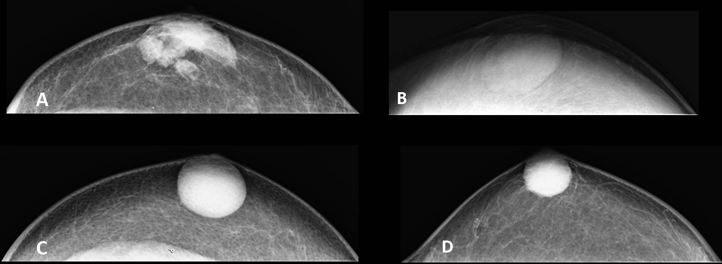
To finish the chapter on male breast disease I am showing mammograms with selected pathology in four different males with palpable retro-areolar lesions. Try to match each case (A-D) with the correct diagnosis.
1. Gynecomastia
2. Carcinoma
3. Epidermoid cyst
4. Simple cyst
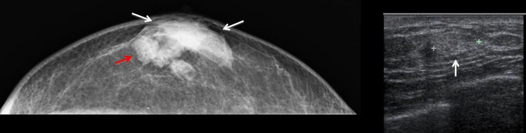
Case A
Case A: the mammogram shows gynecomastia (white arrows) with a nodular area (red arrow) that is solid and hyperechoic on ultrasound examination (arrow). Histologic diagnosis: Papillary carcinoma of the breast
Carcinoma of the male breast has the same imaging characteristics as its female counterpart and accounts for about 1% of all breast carcinomas. Most are infiltrating ductal carcinomas. Papillary carcinomas occur more frequently in males than in females. The diagnosis is suspected when a palpable mass, seen as a solid lesion on ultrasound examination, is found in a male. It is important to note that carcinomas can be hypo- or hyperechoic. Biopsy is mandatory in all males older than 45 years presenting with a solid mammary nodule. Carcinoma of the male breast has no relationship with gynecomastia.
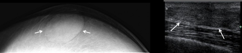
Case B
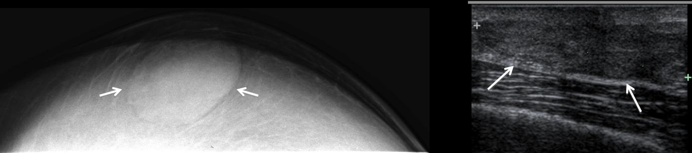
Case B: the mammogram depicts a pseudo-nodular retro-areolar image (arrows). Ultrasound shows a homogenous hypoechoic area (arrows). The findings were identical in the contralateral breast (not shown).
Final diagnosis: Nodular gynecomastia.
Gynecomastia is by far the most common abnormality of the male breast, and usually occurs during puberty. It may be related to the use of certain medications or diseases that increase hormone levels. In most cases, it is idiopathic.
The imaging findings are typical: presence of glandular tissue on mammography and characteristic findings on ultrasound examination. Imaging it is not necessary in pubertal boys, unless it persists.
Gynecomastia does not need treatment. In young people with psychological problems, surgical excision may be indicated if it persists for more than one year.
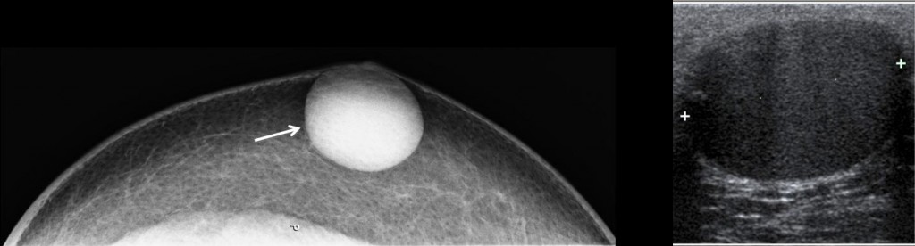
Case C
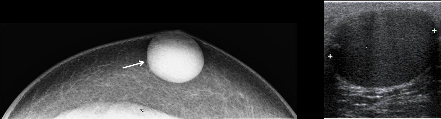
Case C: the mammogram depicts a well-defined rounded lesion (arrow), which is hypoechoic and shows posterior acoustic enhancement on ultrasound examination.
Final diagnosis: epidermoid inclusion cyst
Epidermoid inclusion cysts are not true breast lesions, although they may appear in the breast area. They present as a palpable, slightly painful subcutaneous mass. On mammography, they appear as well-defined rounded peripheral masses. Ultrasound confirms their cystic nature. When infected, they lose these characteristics and behave as a breast abscess.
Epidermoid inclusion cysts can be punctured to relieve the tension, but they can only be cured by surgical removal of the capsule.
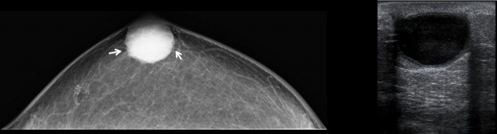
Case D
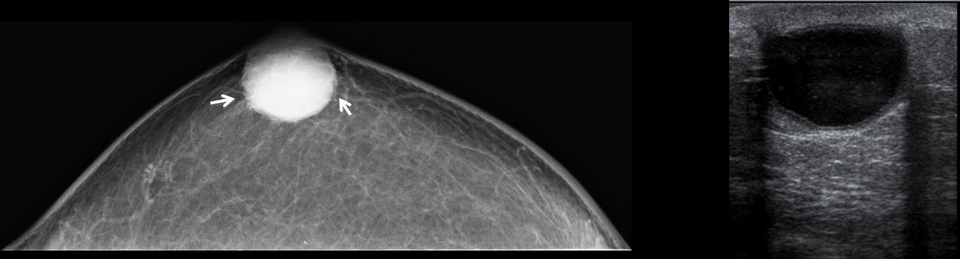
Case D: the mammogram shows a rounded lesion with slight irregularity of the borders (arrows). On ultrasound, a cystic lesion with posterior acoustic enhancement is seen.
Final diagnosis: Simple cyst
Simple breast cysts are uncommon in men. They are usually not as well defined as epidermoid inclusion cysts. The slight irregularity of their margins may raise the possibility of a solid lesion, which should be excluded with US examination (see case 14 of Dr. Pepe’s Diploma).
Ultrasound confirms the cystic nature of the lesion, which can be punctured and evacuated.

Follow Dr. Pepe’s advice
Follow Dr. Pepe’s advice:
- Gynecomastia is the most common abnormality of the male breast.
- A solid breast nodule in an adult male should be investigated to rule out malignancy.
- Male breast carcinomas may be be hypo- or hyperechoic on ultrasound examination.
Recommended reading: Spectrum of disease of the male breast
AJR 2011; 196:W247–W259
Case prepared by Elena Rabanal, MD