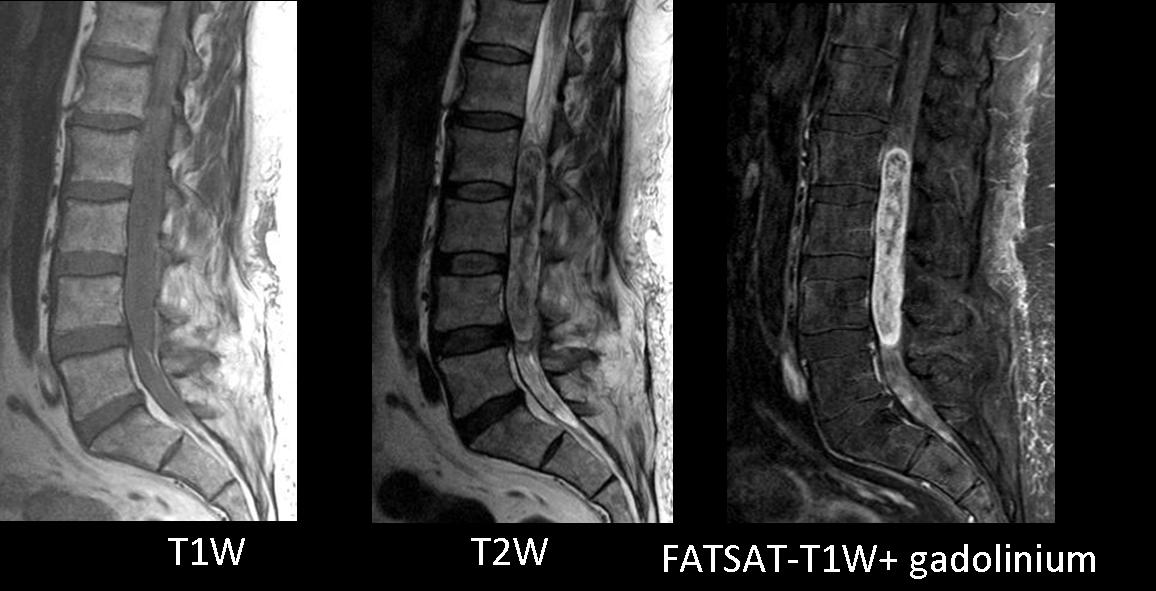
Presenting MRI of lumbar spine in a 57-year-old male with lower-back pain.
1. Ependymoma
2. Meningioma
3. Paraganglioma
4. None of the above
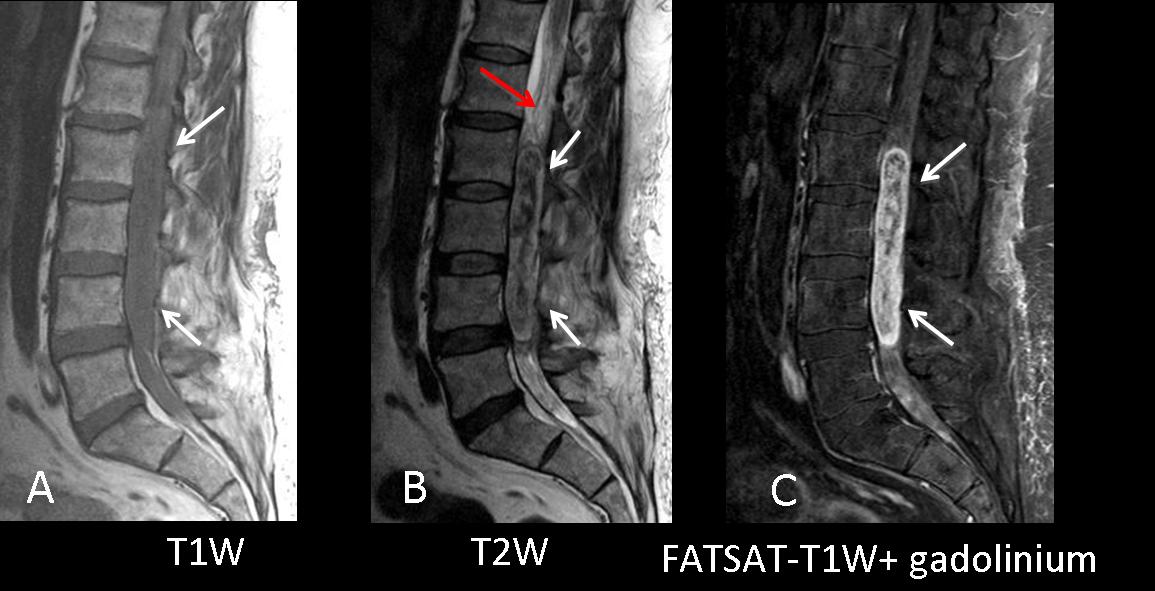
Findings: T1W image shows a huge soft tissue mass (A,B arrows) filling the canal at the lumbar spine. T2W image shows a heterogeneous, mixed signal intensity intradural lesion attached to the filum terminale (B, red arrow). Heterogeneous gadolinium enhancement (C, arrows).
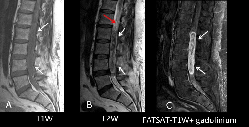
Fig. 1
A mass in the spinal canal in the region of the filum terminale with hypointense central hemorrhagic areas and heterogenous enhancement is highly suggestive of myxopapillary ependymoma.
Final diagnosis: WHO grade I myxopapillary ependymoma.
The differential diagnosis of intradural spinal canal masses should include meningioma, schwannoma, paraganglioma, ependymoma, and intradural metastasis.
Ependymomas account for 40%-60% of primary spinal cord tumours and 90% of primary tumours in the filum terminale. They develop from the ependymal cells lining the central canal of the spinal cord and from ependymal clusters in the filum terminale.
Myxopapillary ependymomas of the filum terminale are usually isointense relative to spinal cord on T1W images and hyperintense on T2W images, with mucin and hemorrhagic content. Flow voids can be seen within the tumour.
Paraganglioma
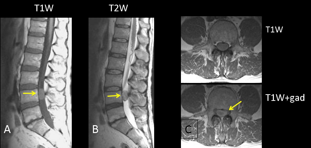
Neoplasm of neuroendocrine origin. Spinal paragangliomas are rare tumours usually located in the cauda equina and filum terminale. Well-circumscribed, hypervascular, intradural masses with intense enhancement, they can present flow voids.
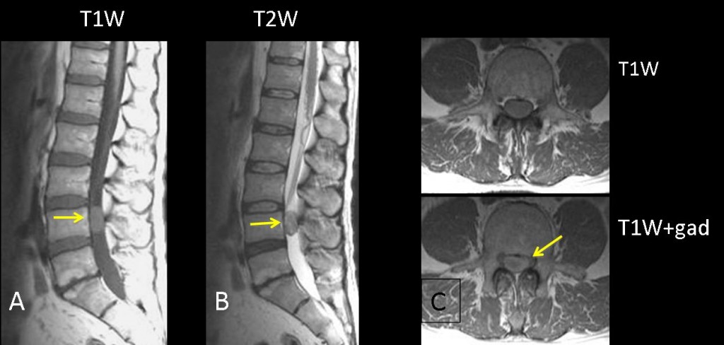
Fig. 2
56-year-old man with back pain and lower extremity paresthesia.
Well-circumscribed intradural mass, isointense relative to spinal cord on T1W and T2W images (A,B arrows). Peripheral enhancement after contrast administration (C, arrow).
Meningioma
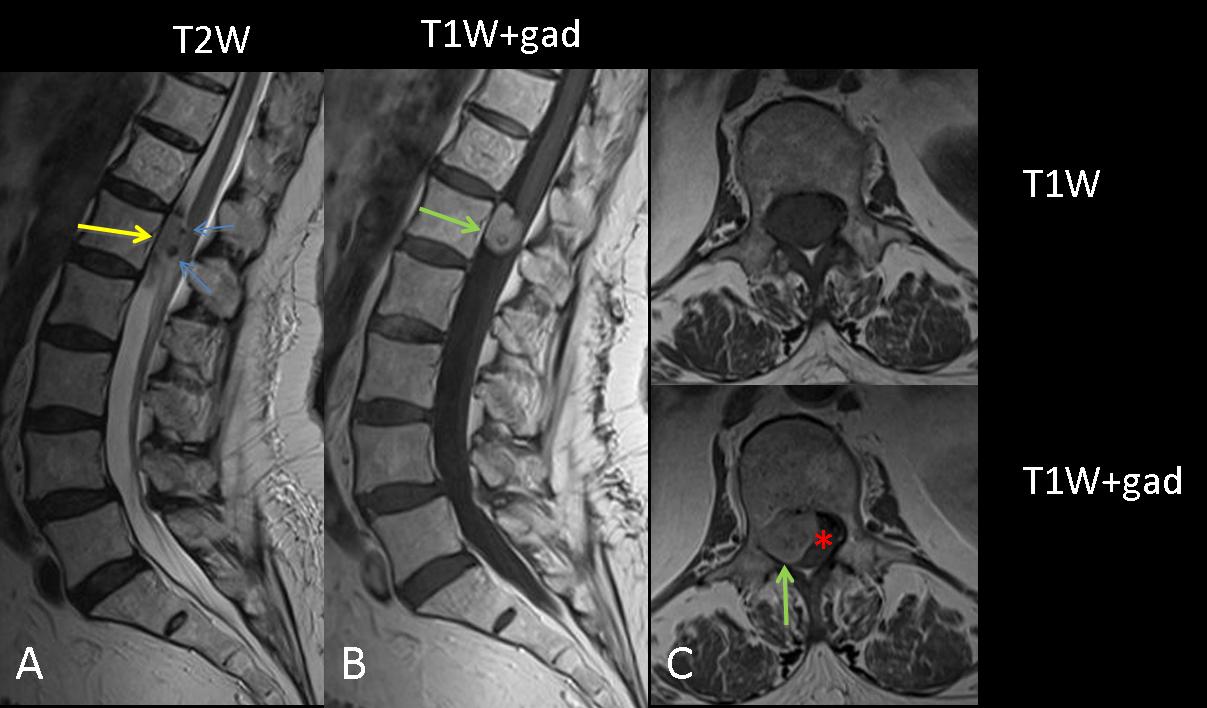
Benign tumours arising from arachnoid. Spinal meningiomas are relatively rare, accounting for 1.2% of all meningiomas of the central nervous system. Isointense with cord on T1 and T2W MR images, showing intense tumoural and dural enhancement, they can calcify.
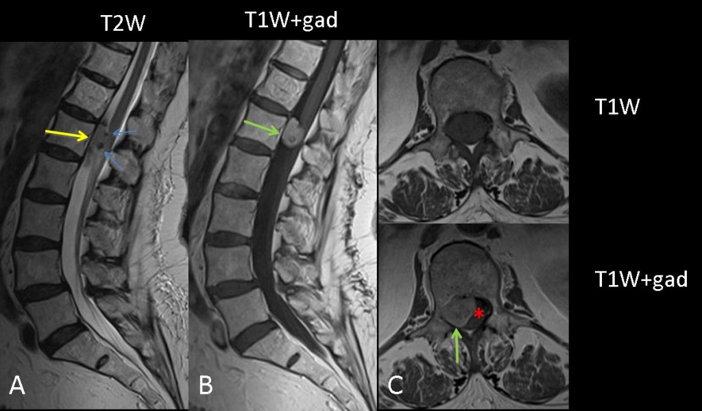
Fig. 3
78-year-old woman with back pain and lower extremity paresthesia. Well-circumscribed intradural mass slightly hyperintense relative to spinal cord on T2W images (A, yellow arrow) with calcifications (blue arrows). Homogeneous enhancement (B,C green arrows) on T1W + gadolinium images. Displacement of the conus medullaris to the left (C, asterisk).

Follow Dr. Pepe’s advice
Follow Dr. Pepe’s advice:
- The differential diagnosis of intradural masses should include ependymoma, paraganglioma, schwannoma, dural metastasis, and meningioma.
- Myxopapillary ependymomas have a predilection for the conus medullaris and filum terminale region, and show mixed signal intensity and heterogenous contrast enhancement.
Recommended reading:
Neoplasms of the spinal cord and filum terminale: radiologic-pathologic correlation. Radiographics 2000; 20:1721-49.
Spinal tumors. European Journal of Radiology 2004; 50:159-176
Case presented by Laura Oleaga, MD