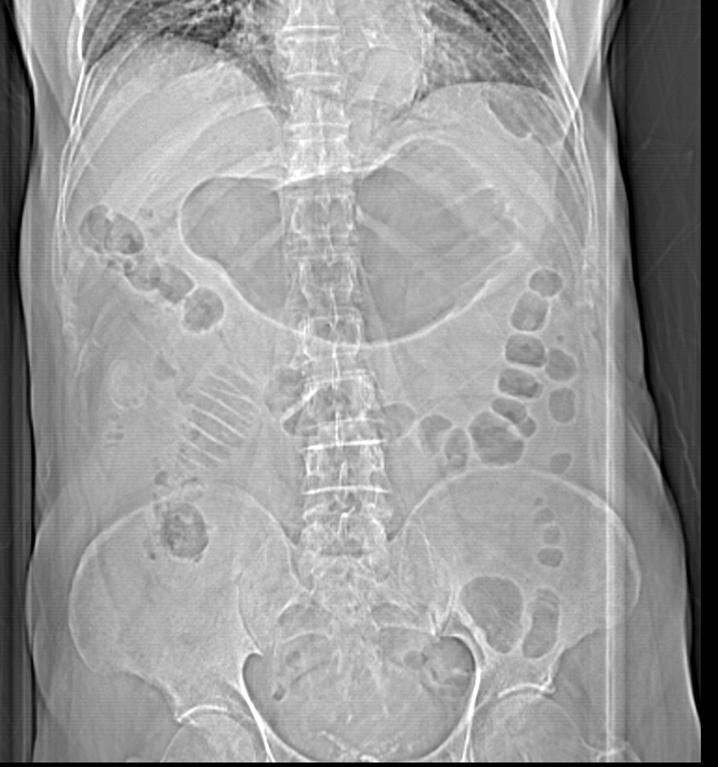
Abdominal films are being displaced nowadays by CT and US; but they are still useful if interpreted properly. In this case you are provided with a supine film of an 84-year-old man who has experienced epigastric pain, vomiting and abdominal distension for several days.
1. Pancreatitis
2. Pyloric obstruction
3. Perforated hollow viscus
4. None of the above
Findings: Dilatation of a small bowel loop and stomach (black arrows). A rounded, laminated calcified structure is present below the right colon (red arrow). A small air bubble is visible superimposed on the liver shadow (yellow arrow).
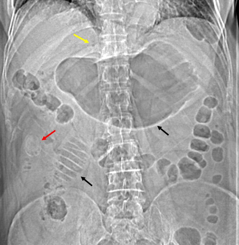
Fig. 2
With the classic radiographic triad: small bowel dilation, possible ectopic gallstone, and suspicion of pneumobilia, the most likely diagnosis is gallstone ileus. CT confirms dilatation of bowel loops, showing an impacted calcified gallstone in the ileum (red arrows). CT also confirms the intrahepatic pneumobilia and distortion of gallbladder anatomy (blue arrows).
Final diagnosis: Gallstone ileus
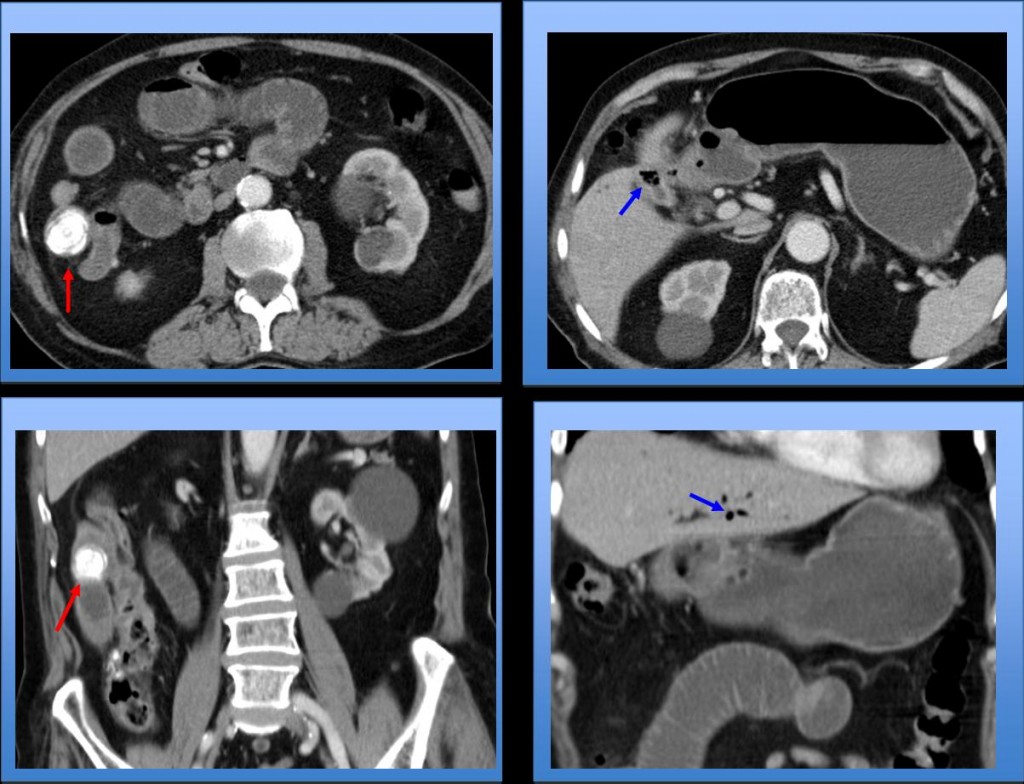
Fig. 3
Gallstone ileus can be suspected in abdominal plain films when a structure that looks like a calcified gallstone is seen outside the normal position of the gallbladder in association with pneumobilia and dilated bowel loops in patients with acute abdomen. Pneumobilia can be missed on plain films if scant air is present. Likewise, the ectopic gallstone might not be seen on plain films if it is poorly calcified or masked by intestinal gas or feces.
CT conclusively confirms gallstone ileus because it always shows the impacted small bowel gallstone, regardless whether or not it is calcified. CT clearly shows the size and exact location of the gallstone.
CT also confirms the other two findings of the classic triad of gallstone ileus: the anatomic distortion of the gallbladder area and the presence of air in the biliary tract, even when scant air is present.
It can be impossible to diagnose gallstone ileus on plain films because the ectopic gallstone and/or the air in the biliary tract may not be visible. In these cases, CT is necessary to reach the diagnosis.
A patient with acute abdomen. In the plain film, only dilated small bowel loops are seen (Fig. 4, white arrows).
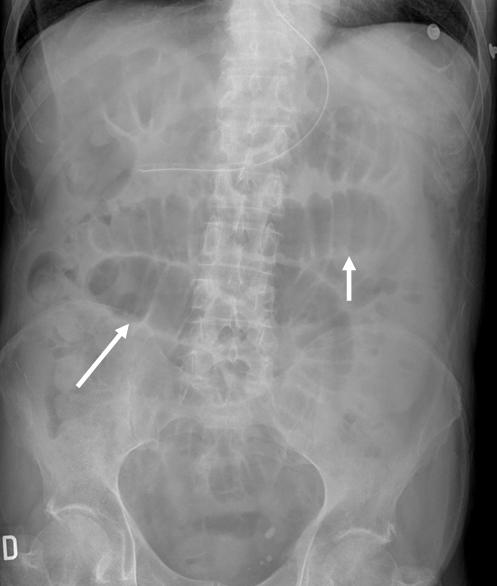
Fig. 4
No ectopic gallstones or pneumobilia are seen. CT confirms an impacted moderatly calcified gallstone in the ileum (Fig. 5, red arrow).
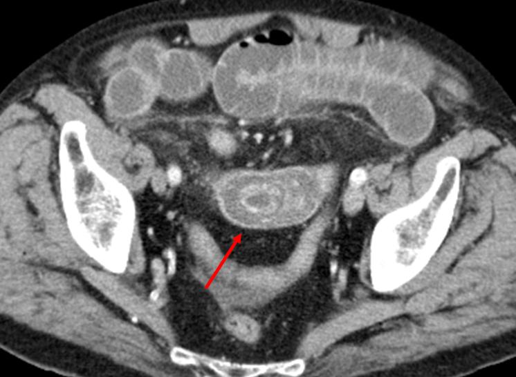
Fig. 5
The differential diagnosis of ectopic calcified gallstone in plain films includes other common abdominal and pelvic calcifications like calcified mesenteric lymph nodes, appendicoliths, or calcified uterine leiomyomas. CT can easily differentiate among these calcifications.
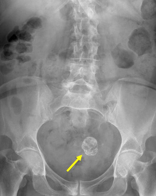
Fig. 6
A rounded calcified image incidentally detected in the pelvis on the plain film is clearly a uterine leiomyoma on CT.
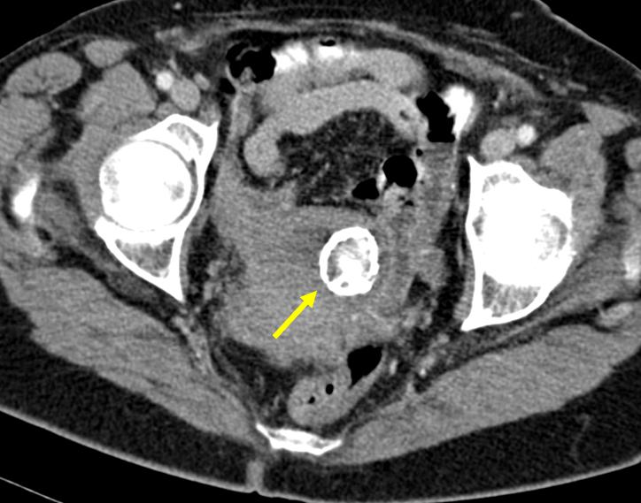
Fig. 7

Follow Dr. Pepe’s advice
Follow Dr. Pepe’s advice:
- Gallstone ileus can be suspected on plain films when the classic triad of intestinal occlusion, ectopic gallstone, and pneumobilia is present.
- In cases in which the ectopic gallstone or pneumobilia are not clearly seen on plain films, the diagnosis of gallstone ileus can be reached only by CT.
Suggested reading:
- Delabrousse E, Bartholomot B, Sohm O, et al. Gallstone ileus: CT findings. Eur Radiol 2000;10(6): 938-40
- Lassandro F, Romano S, Ragozzino A et al. Role of helical CT in Diagnosis of Gallstone ileus and Related Conditions. AJR 2005;185: 1156-1165
Case prepared by Julio Martin MD