Today, I want to show you a classic case of vertebral affectation. I’m hoping for 100% correct answers.
59-year-old female with lumbar pain.
1. Metastases
2. Lymphoma
3. Paget’s disease
4. All of the above
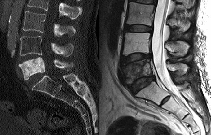
Fig 3
Findings:
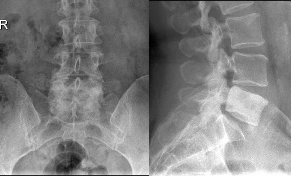
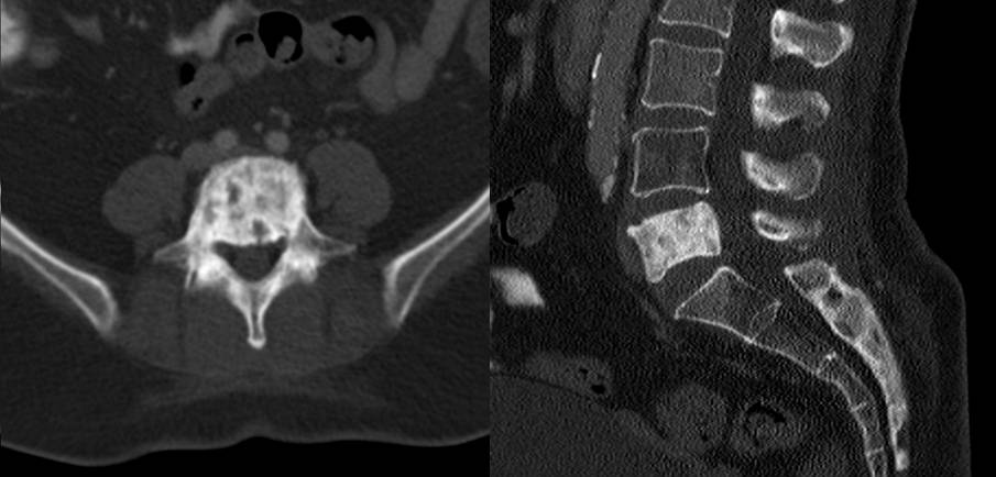
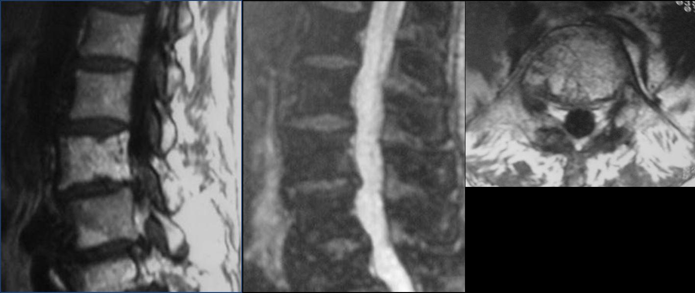
Increased density of the L5 vertebral body, without loss of volume. There is no disc involvement.
Neither CT nor MRI show a soft tissue mass. �Given the homogeneity and the absence of soft tissue mass, the most likely diagnosis is metastases.
Final diagnosis: metastases from breast carcinoma
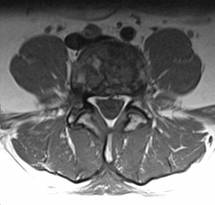
Fig 4
The solitary dense vertebra (ivory vertebrae) is a finding relatively easy to detect due to the increased opacity compared to other vertebrae. �The differential diagnosis is relatively simple and includes metastases (prostate in the male and breast in the female), Paget’s disease, lymphoma, and reactive sclerosis.
In Paget’s disease, the vertebra shows a picture frame appearance, caused by cortical thickening and coarse trabeculation. There is usually an increase in the size of the vertebra, highly suggestive of Paget’s disease (Fig. 5).
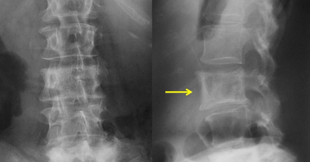
Fig. 5. Paget’s disease. Note the picture-frame aspect of L-3 and the increase in size compared to the vertebras above and below.
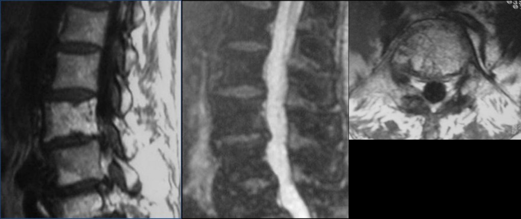
Fig. 6
Unlike the low signal intensity of metastatic vertebrae in T1W images, the vertebra of Paget’s disease contain large areas of fat tissue.
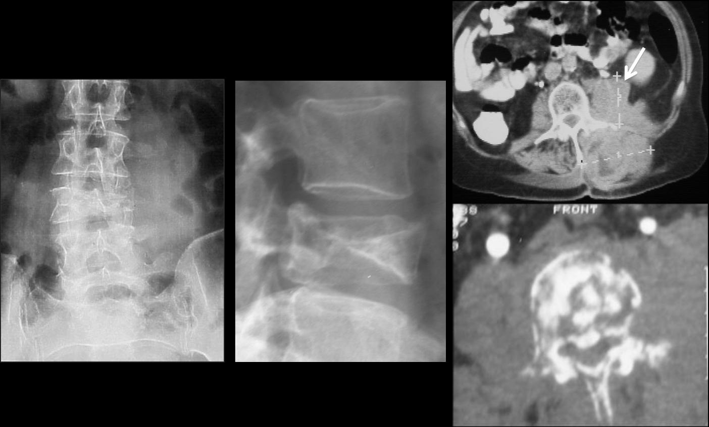
Lymphoma causes increased bone density of the affected vertebrae, but it is usually accompanied by a destructive pattern and large soft tissue mass (Fig. 7).
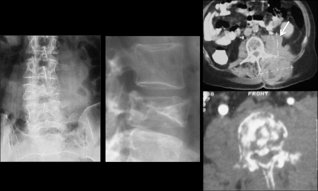
Fig. 7. Lymphoma of L3. Note the destructive pattern of the bone and the soft tissue mass on CT (arrow).
The lack of fragments protruding anteriorly or posteriorly usually rules out posttraumatic lesions. The absence of disc involvement would go against infectious involvement.
The most reliable finding is the presence of soft tissue mass. Bone involvement can be both homogeneous and heterogeneous.
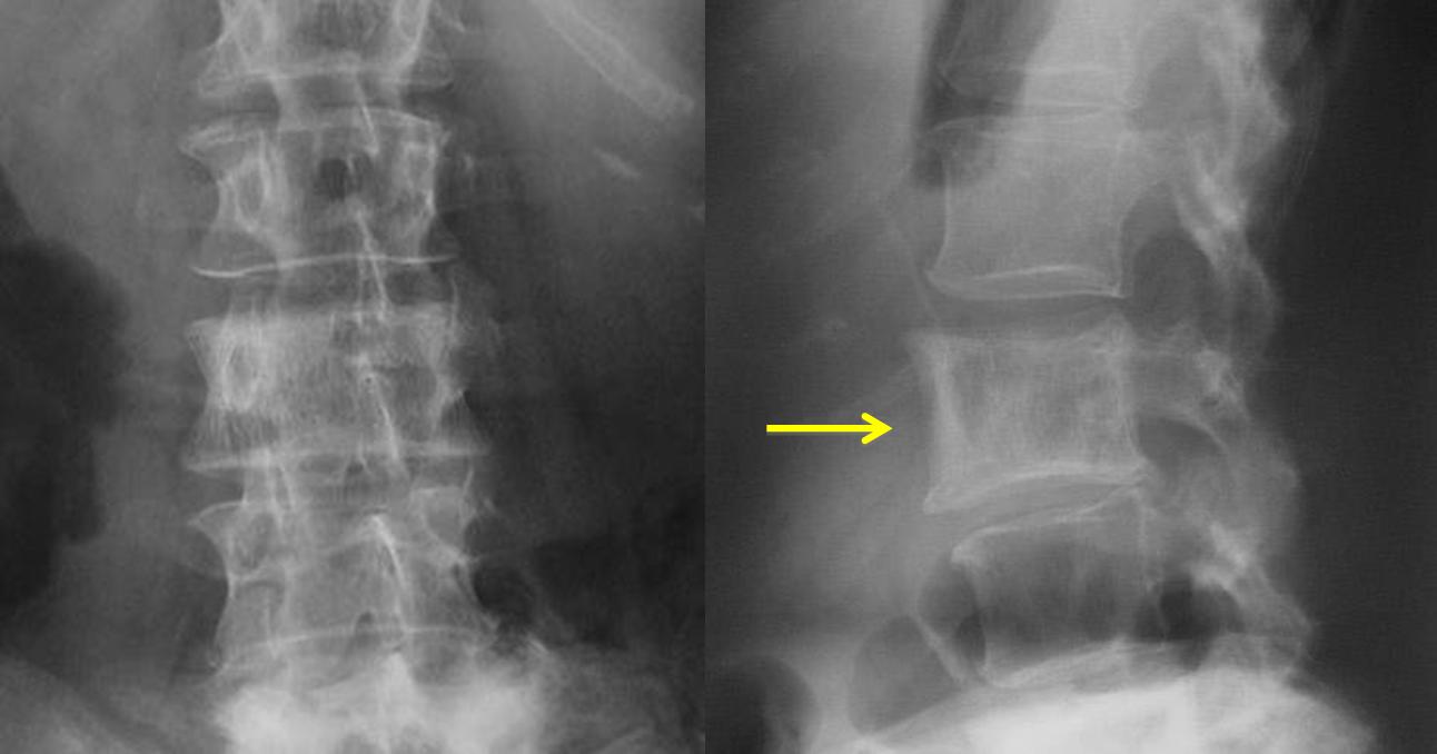
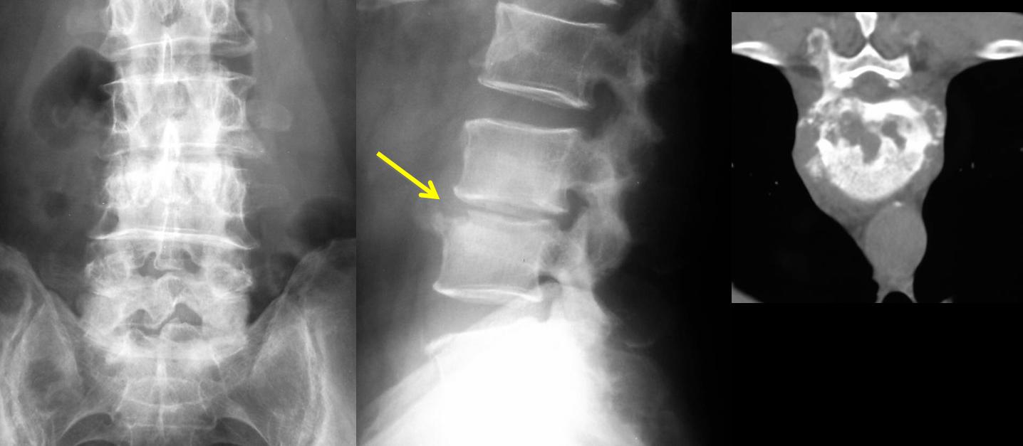
Reactive sclerosis may be due to inflammatory, traumatic or degenerative disease. Rarely involves a single vertebra and the disc is usually narrowed (Fig. 8).
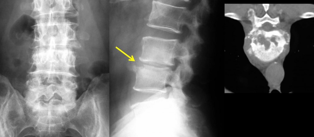
Fig. 8. 52-year-old male with lumbar pain lasting 6 months. Note the sclerosis of L3-L4 and the narrowing of the disc space (arrow) secondary to Brucella infection.

Follow Dr. Pepe’s advice
Follow Dr. Pepe’s advice:
- Increased density of a vertebral body (ivory vertebra) can be detected easily on plain films.
- It is important to determine the homogeneity of the damage, the trabecular pattern, the existence of soft tissue mass or disc affectation to reach an appropriate diagnosis.
- TC and MRI are very useful for detecting these alterations.
Recommended reading: TS Graham. The Ivory Vertebra Sign. Radiology 2005; 235:614–615
Case prepared by Dámaso Aquerreta MD