Dr. Pepe’s Diploma Casebook: Case 54 – SOLVED!
Dear Friends,
Welcome to this week’s case. Radiographs belong to a 43-year-old male with chest pain. Leave me your thoughts and diagnosis in the comments and come back on Friday for the answer.
Diagnosis:
1. Hypertrophic cardiomyopathy
2. Mitral disease
3. Non-cyanotic congenital heart disease.
4. None of the above
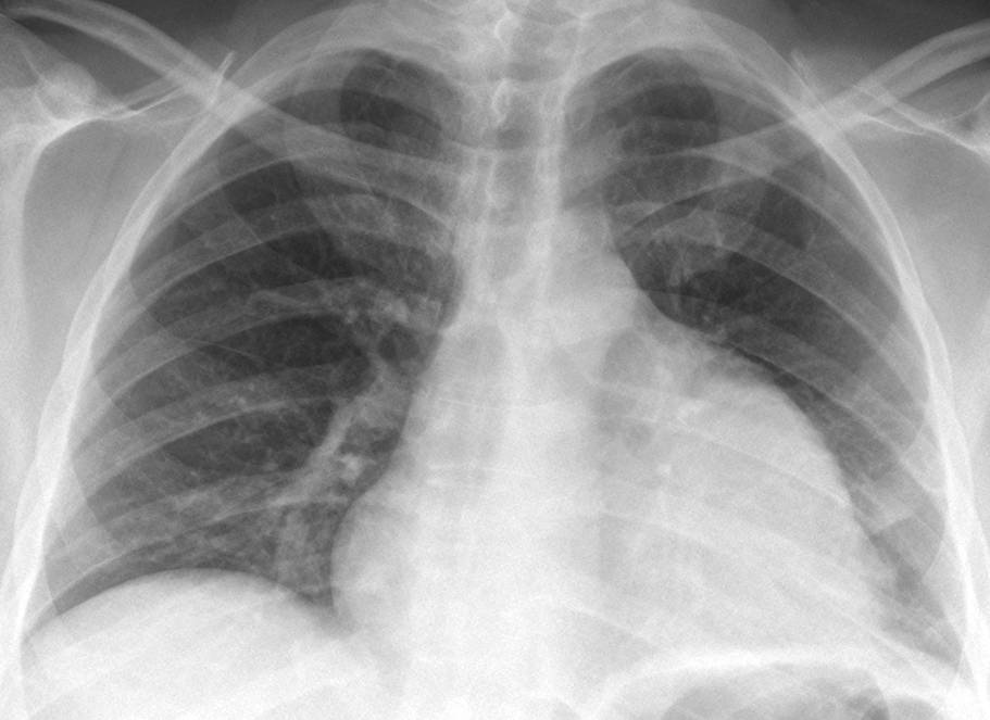
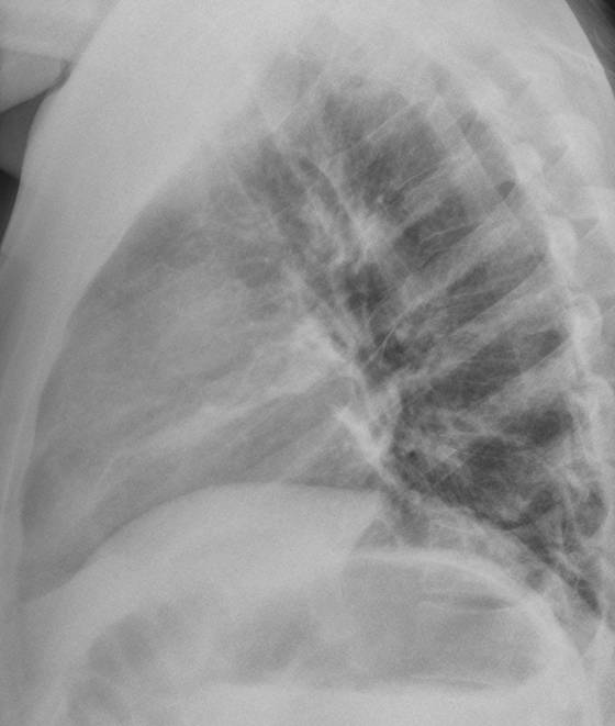
Findings: radiographs show an enlarged cardiac silhouette with abnormal contour of the left heart border (A, arrows). Mitral disease can be ruled out (no left atrial enlargement or vascular redistribution). There are no signs of right-to-left shunt, which is the hallmark of non-cyanotic heart disease. Hypertrophic cardiomyopathy does not change the heart contour until the late stages. A clue to the diagnosis is the faint rounded opacity in the lateral view (B, arrows), which prompted a CT study.
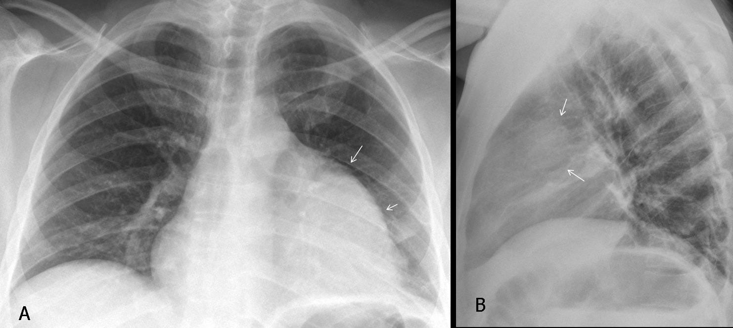
Fig. 1
Enhanced CT demonstrates that the prominent left heart border is due to a large, poorly enhancing mass apposed to the left aspect of the heart (A,B, arrows). Biopsy of the mass diagnosed a mediastinal sarcoma.
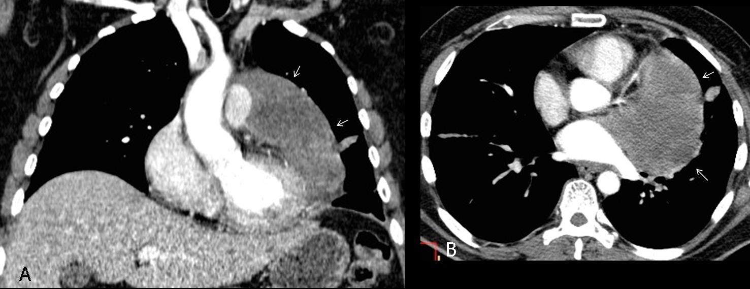
Fig. 2
Final diagnosis: Mediastinal sarcoma simulating cardiomegaly.
The presentation today focuses on causes of false cardiomegaly. When the cardiac silhouette is seen to be greater than or equal to 50% the diameter of the chest, our immediate response is a diagnosis of cardiomegaly, and most of the time we are right. But, occasionally, the apparent cardiomegaly is not real and is caused by a process that simulates it.
One uncommon cause of false cardiomegaly is the presence of a lower anterior mediastinal mass apposed to the heart and having a similar density, thus creating the illusion of an enlarged cardiac silhouette. The diagnosis is suspected based on a lack of clinical and EKG signs of heart disease and an abnormal appearance of the cardiac contour. Confirmation of the suspected diagnosis is easily made with CT.
Any anterior mediastinal tumour can lead to this misdiagnosis. The most common ones are of thymic origin because they have a tendency to descend and locate around the heart (Fig. 3). Thymolipoma is an unusual fatty tumour that surrounds the heart and simulates overall cardiomegaly (Fig. 4).
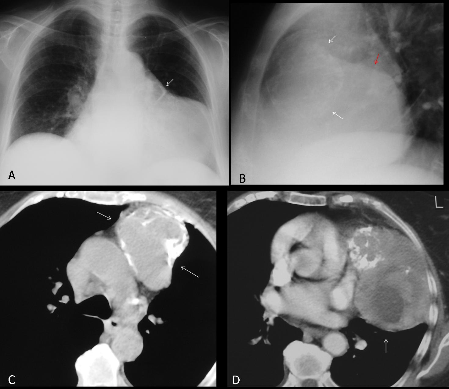
Fig. 3
Fig. 3 (above): 58-year-old man with apparent cardiomegaly. Nonetheless, a diagnosis of mediastinal mass is suggested by the extra-cardiac calcification (A,B white arrow) and a double contour in the lateral film (B, red arrow). Axial CT confirms the partially calcified mediastinal mass (C,D, arrows), apposed to the left heart border. Diagnosis: Malignant thymoma.
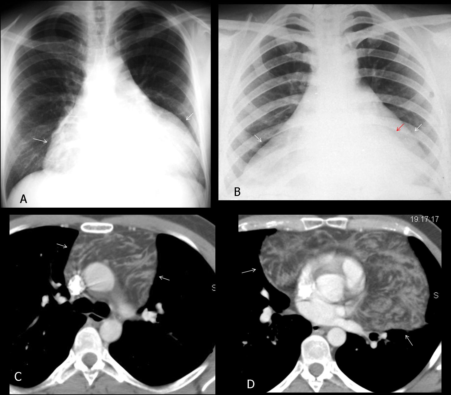
Fig. 4
Fig. 4 (above): two patients with thymolipoma. In both cases, the tumour surrounds the heart, simulating overall cardiac enlargement (A, B, white arrows). In B, the outline of the heart is faintly seen (B, red arrow), enclosed by tumoural fat. Axial CT in the first patient confirms the fatty nature of the thymolipoma surrounding the heart (C,D, arrows)
By far the most common cause of false cardiomegaly is pericardial effusion of any cause. In the PA chest film, it is typically seen as a globular cardiac silhouette with decreased pulmonary circulation. However, this appearance may be deceiving because cardiomyopathy can show similar features (Fig. 5). In my experience, the best sign of pericardial effusion is posterior displacement of the epicardial fat in the lateral view (fat pad sign) (Fig. 6). On visualisation of the anterior mediastinal fat, pericardial thickness can be measured (Fig. 7), and this should not exceed 2-3mm in normal conditions. The fat pad sign is not always seen, but when present, it is a highly reliable finding. Nonetheless, it is not pathognomonic and should always be followed by echocardiography to confirm the suspected effusion.
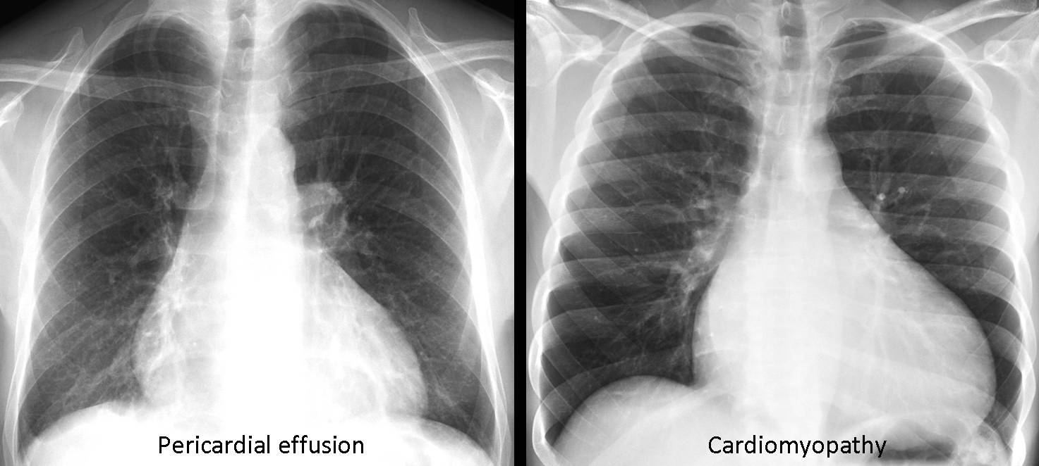
Fig. 5
Fig. 5 (above): two patients; one with pericardial effusion (A) and the other with cardiomyopathy (B). Note the similar appearance of these conditions.
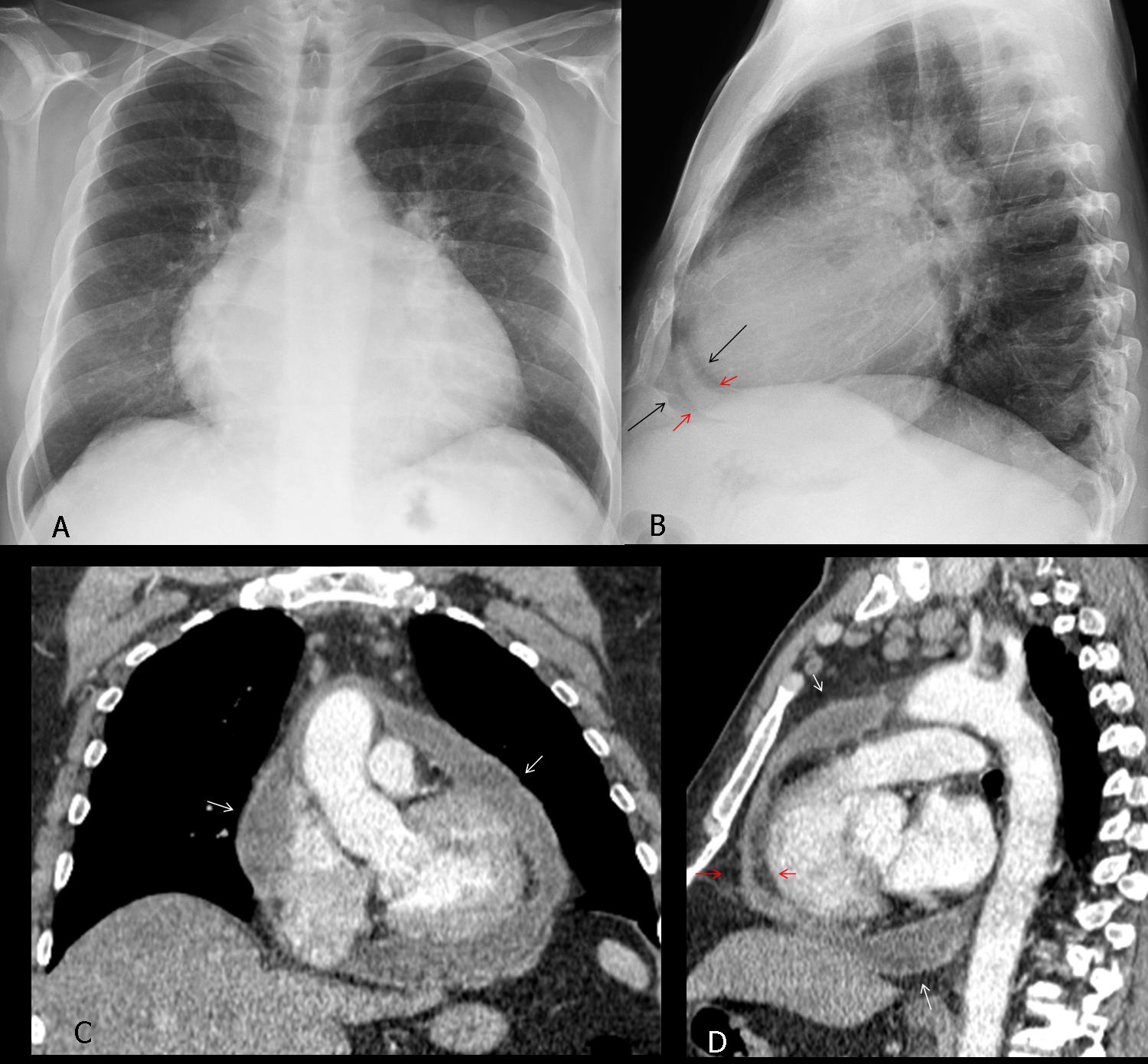
Fig. 6
Fig. 6 (above): 46-year-old man with liver cirrhosis and pericardial effusion. Note the typical globular appearance of the cardiac silhouette in the PA view (A). The diagnosis is evident in the lateral view, which shows the thickened pericardium (B, red arrows) between the epicardial and mediastinal fat (black arrows). Coronal and sagittal CT confirm the presence of a moderate amount of pericardial fluid (C,D, white arrows). Note the fluid between the epicardial and mediastinal fat in the lateral view (D, red arrows).
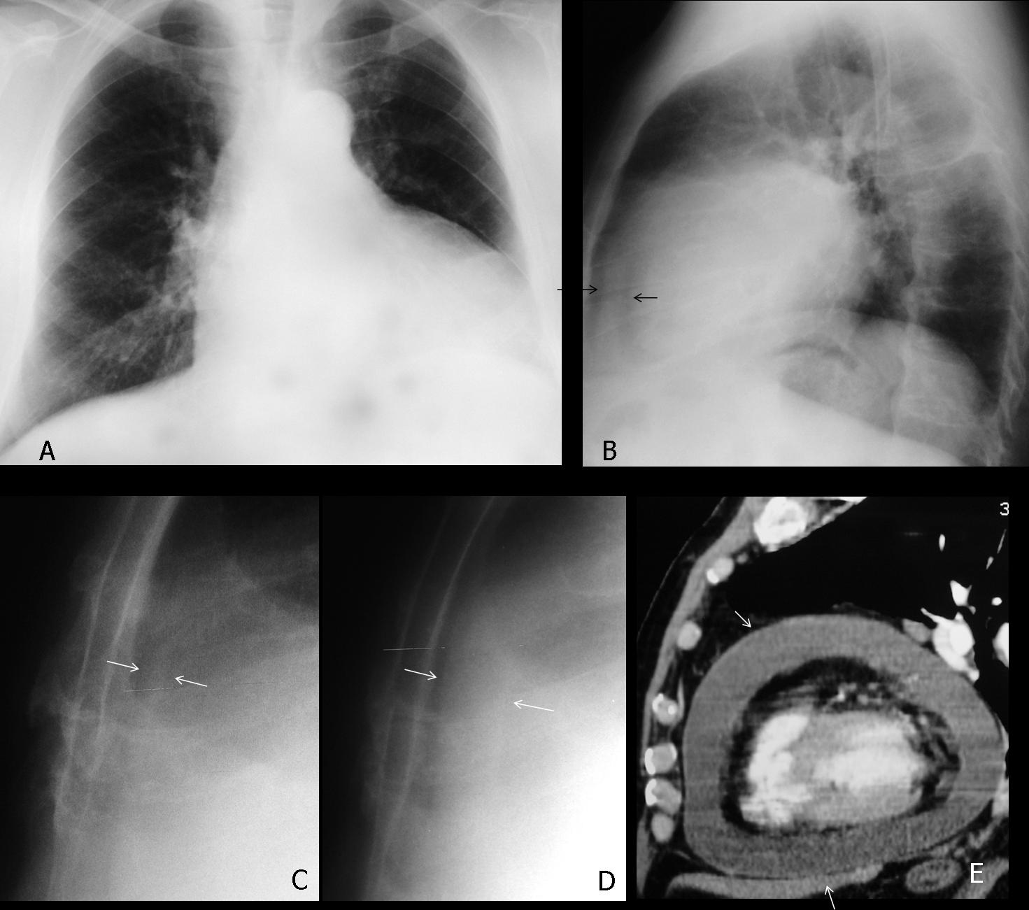
Fig. 7
Fig. 7 (above): 53-year-old man with pericardial effusion simulating cardiomegaly in the PA view (A). The diagnosis is suggested in the lateral view by the thickened pericardium between the epicardial and mediastinal fat (B, arrows). Cone down lateral view taken three months earlier shows normal pericardium (C, arrows). Compare with the present film (D, arrows). Sagittal CT confirms the presence of a large pericardial effusion (E, arrows).
Pericardial metastasis is not rare in patients with disseminated malignant disease. The most common primaries are lung and breast. They can be suspected by the irregular contour of the cardiac silhouette and confirmed with CT (Fig. 8).
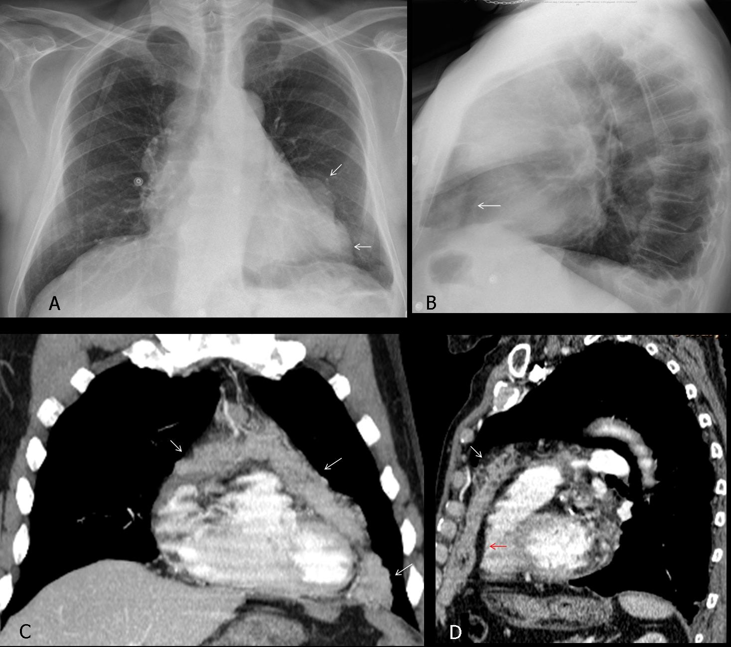
Fig. 8
Fig. 8 (above): patient with widespread malignancy. Pericardial metastasis is suggested by the abnormal heart contour (A, arrows) and epicardial fat displacement in the lateral view (B, arrow). Axial and coronal CT images confirm metastasic invasion of pericardium by solid masses (C,D, white arrows). Note the displaced epicardial fat in the lateral view (D, red arrow).
In young people, another cause of apparent cardiomegaly is severe pectus excavatum. The sternum compresses and displaces the heart towards the left, simulating cardiac enlargement (Fig. 9). The diagnosis is readily apparent by looking at the lateral film.
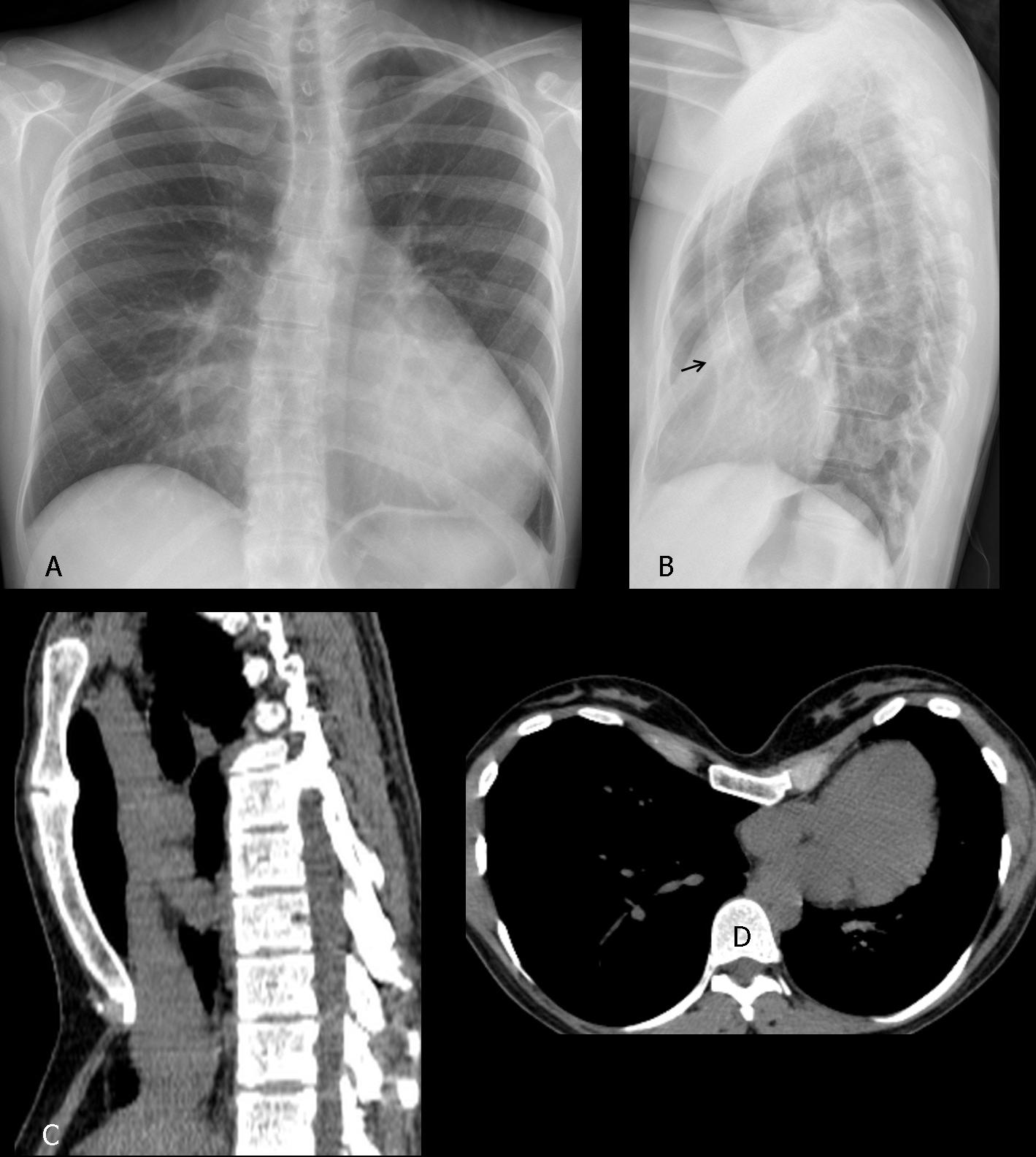
Fig. 9
Fig. 9 (above): 17-year-old male with marked pectus excavatum simulating cardiomegaly in the PA chest film (A). Lateral film confirms the pectus (B, arrow). Sagittal and axial CT images (C,D) confirm marked narrowing of AP diameter of the chest and displacement of the heart towards the left.
Last, but not least, remember that heart size cannot be evaluated properly in expiratory and supine films (Fig. 10).
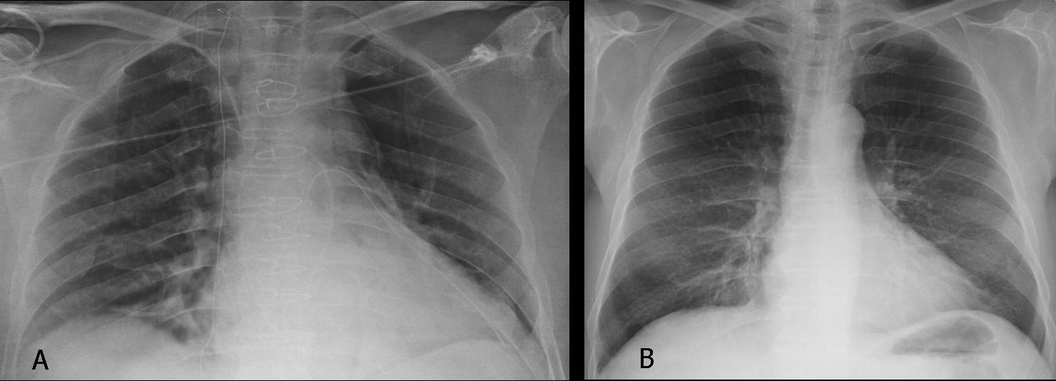
Fig. 10
Fig. 10 (above): supine film (A) in which the cardiac silhouette appears enlarged (A). Previous upright film in the same patient shows a normal-sized heart (B).

Follow Dr. Pepe’s advice
Follow Dr. Pepe’s advice:
1. The most common cause of false cardiomegaly is pericardial effusion.
2. The most reliable sign of pericardial effusion is posterior epicardial fat displacement in the lateral view.
3. An uncommon cause of false cardiomegaly is an anterior/inferior mediastinal mass.
4. In an asymptomatic young person with enlarged heart, think of pectus excavatum.


