Dr. Pepe’s Diploma Casebook: Case 60 – SOLVED!
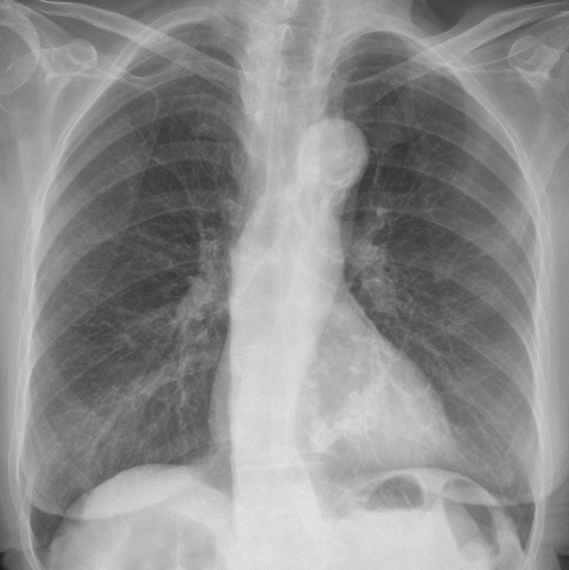
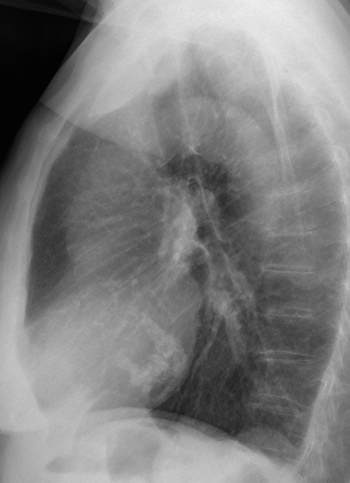
Today I am showing chest radiographs of a 78-year-old woman with moderate dyspnoea. Check the images below, leave your thoughts and diagnosis in the comments, and come back on Friday for the answer.
Diagnosis:
1. Mitral valve calcification
2. Pericardial calcification
3. Calcified ventricular aneurysm
4. None of the above
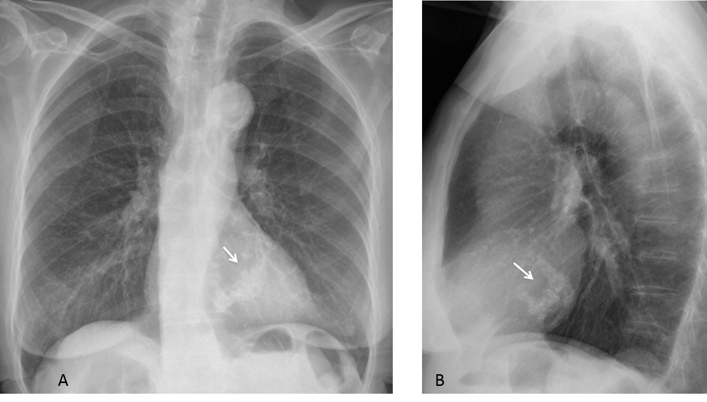
Findings: PA and lateral radiographs show a coarse reverse C-shaped calcification projected over the cardiac silhouette (A,B arrows). The appearance and location are practically pathognomonic of calcification of the mitral annulus.
Final diagnosis: calcified annulus fibrosus of the mitral valve.
This case is presented to discuss cardiac calcifications in the chest radiograph. A simple classification divides them into those projected over the cardiac shadow (central calcifications) and those outlining the cardiac silhouette (peripheral calcifications). The first ones are mainly related to the aortic and mitral valves and the second to calcium in the myocardium or pericardium.
As you will see during this presentation, the lateral chest film plays a crucial role in discovering and characterising cardiac calcifications.
CENTRAL CALCIFICATIONS. In my experience, the most common occur in the annulus fibrosus of the mitral valve (Fig. 2), resulting from a degenerative process that mainly affects the elderly. It is usually asymptomatic, but a small percentage of patients may have moderate mitral regurgitation, secondary to malfunctioning cordae tendinae. As was mentioned earlier, the radiographic appearance is practically pathognomonic. It should not be confused with a calcified mitral valve.
Fig. 2 (above): mitral annulus calcification. Note the typical reverse C shape (A,B, arrows). CT confirms the appearance and location (C-E, arrows). The aortic valve also shows calcification (B,E, red arrows).
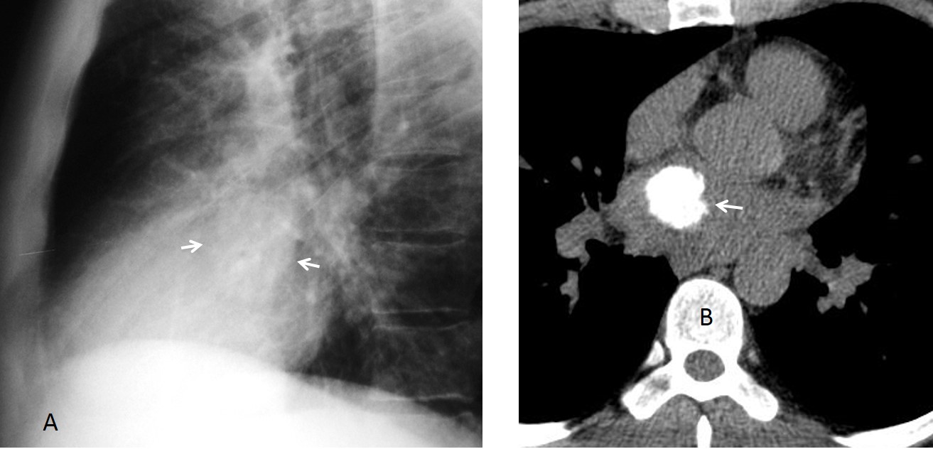
A variant of annulus calcification is the so-called caseous necrosis of the mitral annulus. In this condition, the calcified rim surrounds a toothpaste-like substance. The plain film shows a rounded calcification in the area of the annulus (Fig. 3 A,B), whereas CT depicts a rim calcification with a low-density center (Fig. 3 C,D). It is important to be aware of this variant, so it is not mistaken for a significant pathology.
Fig. 3 (above): caseous necrosis of the annulus. Note the rounded calcification in the region of the annulus (A,B, arrows). Coronal and sagittal CT show a calcified rim with a less dense center (C,D, arrows). The appearance is typical of caseous necrosis of the annulus and should not be mistaken for a tumour or infection.
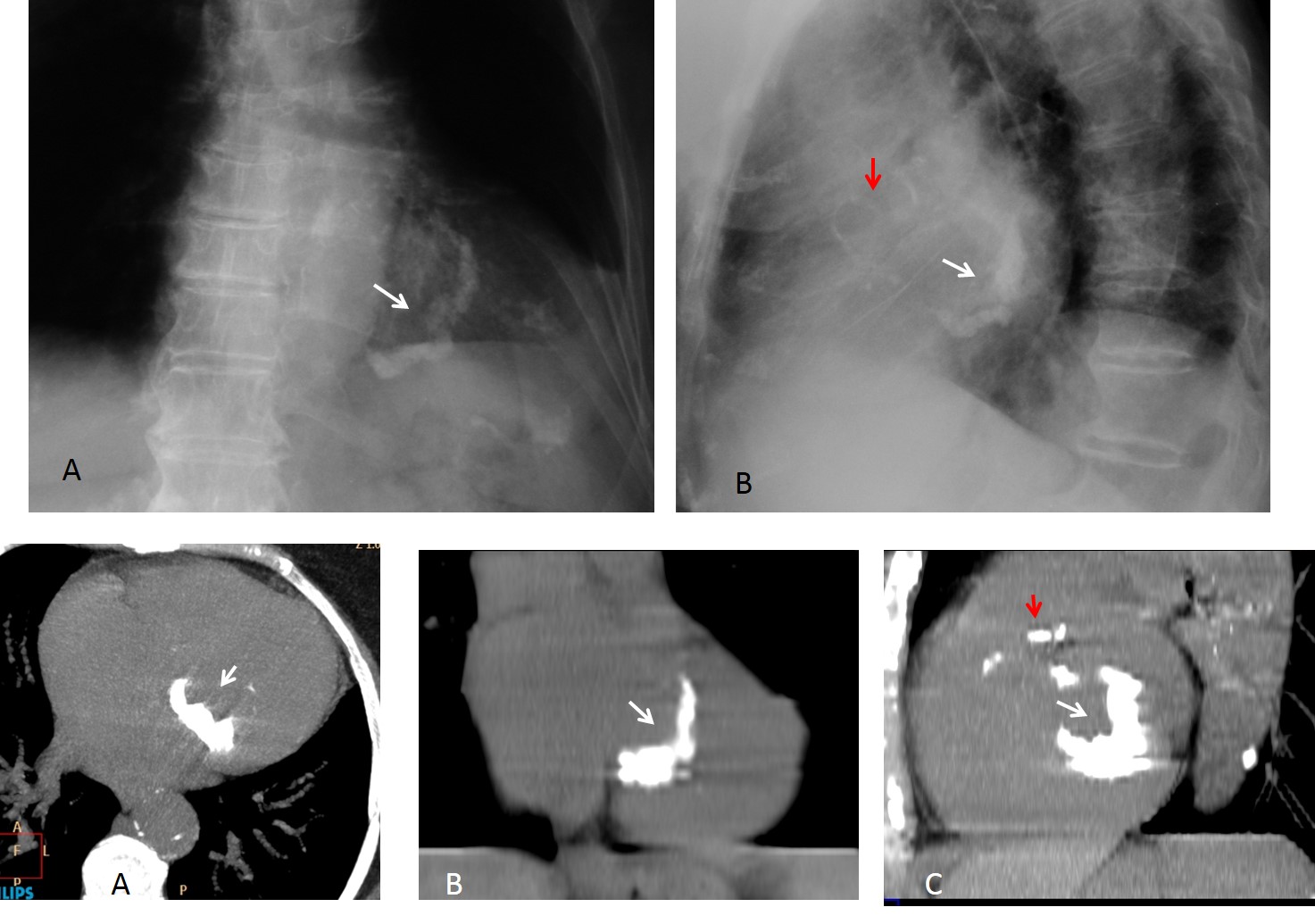
Calcified aortic valve is the second most common type of central calcification. It is better seen in the lateral view as coarse calcium in the area of the aortic root (Fig. 4 A) and is easily confirmed with CT or fluoroscopy (Fig. 4 B,C ). Clinical significance depends on the age of the patient. In reasonably young patients (around 40 years old), this finding is highly suspicious for a bicuspid valve with aortic stenosis. In elderly people, it is usually less significant.
Fig. 4 (above): calcification of the aortic valve, better seen in the lateral view in the area of the aortic root (A, arrow). Sagittal and coronal CT confirm the location of the calcium at the aortic valve (B,C, arrows)
Calcification of the mitral valve is seen in a position below the aortic valve in the lateral view (Fig. 5). This finding was more common in the past, when rheumatic fever was prevalent. Currently, rheumatic heart disease has nearly disappeared from the Western world. For this reason, mitral valve calcification is rare nowadays, although it is still seen in individuals from less privileged countries.
Fig. 5 (above): lateral chest film shows calcium in the mitral valve (A, arrow). Sagittal and coronal CT confirm the location (B,C arrows).
PERIPHERAL CALCIFICATIONS are located in the heart’s outline. They are usually due to myocardial or pericardial disease.
Myocardial calcification is nearly always secondary to an aneurysm following infarction. It is seen as a thin line that curves inwards, usually located at the left ventricle apex (Fig. 6). A small percentage are incidental findings, resulting from a previous silent infarction (Fig. 7).
Fig. 6 (above): 65-year-old man with myocardial infarction and calcified aneurysm of the left ventricle. Note the thin curvilinear calcification at the apex of the heart (A,B, arrows).
Fig. 7 (above): calcified ventricular aneurysm found incidentally in a 54-year-old man (A, arrows). Unenhanced and enhanced axial CT confirm the aneurysm (B,C, arrows), which has a calcified mural thrombus (B,C, red arrow).
Pericardial calcification is a sequela of previous pericarditis due to infection, trauma, or cardiac surgery. Its presence suggests constrictive pericarditis, although this is not always the case and the condition must confirmed clinically. The calcification is usually coarse and is not limited to a specific area (Figs. 8,9).
Fig. 8 (above): sequela of viral pericarditis in a 57-year-old man. Note the coarse peripheral calcification around the heart (A,B, arrows). The patient did not have symptoms of constrictive pericarditis.
Fig. 9 (above): calcified tuberculous pericarditis. The PA view shows a rounded peripheral calcification (A, arrow), which simulates a ventricular aneurysm. The lateral view depicts extensive pericardial calcification (B, arrows). Note the apical lung changes secondary to the previous TB.
Coronary calcifications are poorly seen in the chest radiograph; CT is the technique of choice for their study. When visible in the plain film, they appear as parallel tracts arising from the aortic root (Fig. 10 A). It is important not to confuse them with stents, which are sharply defined and far more common nowadays (Fig. 10 B).
Fig. 10 (above): railroad track calcifications (A, arrows) typical of calcium in the coronary artery. Do not mistake these features for coronary stents (B, arrow).
For the sake of completeness, cardiac tumours should also be mentioned. Primary cardiac neoplasms are uncommon and are usually discovered because of cardiac symptoms or embolic phenomena. When the tumor is calcified, the calcium is barely visible in plain films (Fig. 11). Left atrial myxomas are the most common of these tumours.
Fig. 11 (above): primary cardiac osteosarcoma, discovered because of tumor emboli to the brain. Even though it is highly calcified, the tumor is barely visible in the lateral chest film (A, arrows). Unenhanced CT shows the calcified tumor in the left atrium (B, arrow).
Follow Dr.Pepe’s Advice:
1. The lateral view is very helpful for detecting and categorizing cardiac calcifications.
2. Central calcifications usually occur at the mitral annulus and aortic valve.
3. Myocardial calcification usually locates at the apex of the heart. Pericardial calcification can occur anywhere on the periphery of the heart.
4. Visible stents are more common than calcified coronary arteries.





Mitral valve calcification
What a great case!!! Even though i have never seen a calcified ventricular aneurysm on X-ray, i believe that is it. But i am worried about right lower lobe where seems to exist a lesion most prominent on profile image.
This soft-tissue opacity lesion seen posteriorly on lateral image is a small Bochdalek hernia, probably…
Yes, it is
Don’t worry about RLL. On Friday i will show you several types of calcifications
Mitral annular calcification
I agree
Calcificación severa anular Mitral. Me mosquea no ver un claro crecimiento auricular izquierdo.
La calcificación del anillo mitral no entraña aumento de tamaño de la aurícula.
Answer 4.
2. Pericardial calcification
Mitral annular calcification
Mitral valve (coarse, C-shaped) calcification . No signs of LV enlargement and LV aneurysm would be located anteriorly.
Left posterior hemidiaphragm bulge (like in case 20) – needs further examination.
She is 78. probably a small Bochladek hernia
1. Mitral valve calcification
Mostly 3: aneurysmal calcification
Dear Sir greetings..It looks like Mitral valvular calcification with Aortic knuckle calcification and changes of osteopenia
Looks like dense C-shape mitral annular calcification. Some nodularity to it makes me think there might be some focal superimposed caseous necrosis.
…per le opzioni 2 e3 vi deve essere il raccordo anamnestico di una pregressa pericardite( con calcificazione più”sottile” ed a ” guscio”) o di un infarto del miocardio….una calcificazione di un aneurisma della coronarica,avrebbe un’ altra sede … Invece per forma e sede e’ più’ probabile che rappresenti l’esito di una necrosi caseosa , con calcificazione grossolana dell’ anulus , di una valvola mitralica.
Could it be liquefaction necrosis of mitral annulus?
As far as I know, the appearance of casseous necrosis in the plain film has not been described in the literature.
I have collected six cases (which makes me the world’ expert 😉 ) and all of them have a rounded appearance, instead of being C-shaped.
Calcified Ventricular aneurysmal
Mitral valve calcification
…..errata corrige : Genchi bari italia( non granchi)
….dovresti spiegarci perché’ la necrosi caseosa e successiva calcificazione interessa solo la valvola mitrale e non le altre valvole cardiache?Grazie…
I don´t know. May be due to the anatomy of the chordae tendinae of the mitral valve. There are several articles about this condition. Will look it up and get back to you.
At this time of the week I believe it is correct to give the right answer: the appearance is practically pathognomonic of calcified mitral annulus.
Congratulations to Mohammad Alshafie, who was the first to mention the right diagnosis.
Dr.Pepe, wasn’t me to mention the diagnosis earliest !
Sorry Dr.Pepe I should have answered Mitral annulus….
Sorry, as you say, it is not the same mitral valve than mitral annulus. Still, the aim of the cases is to clarify concepts and, hopefully, to enjoy them. It is not a matter of winning or losing.
Mitral valve calcification.