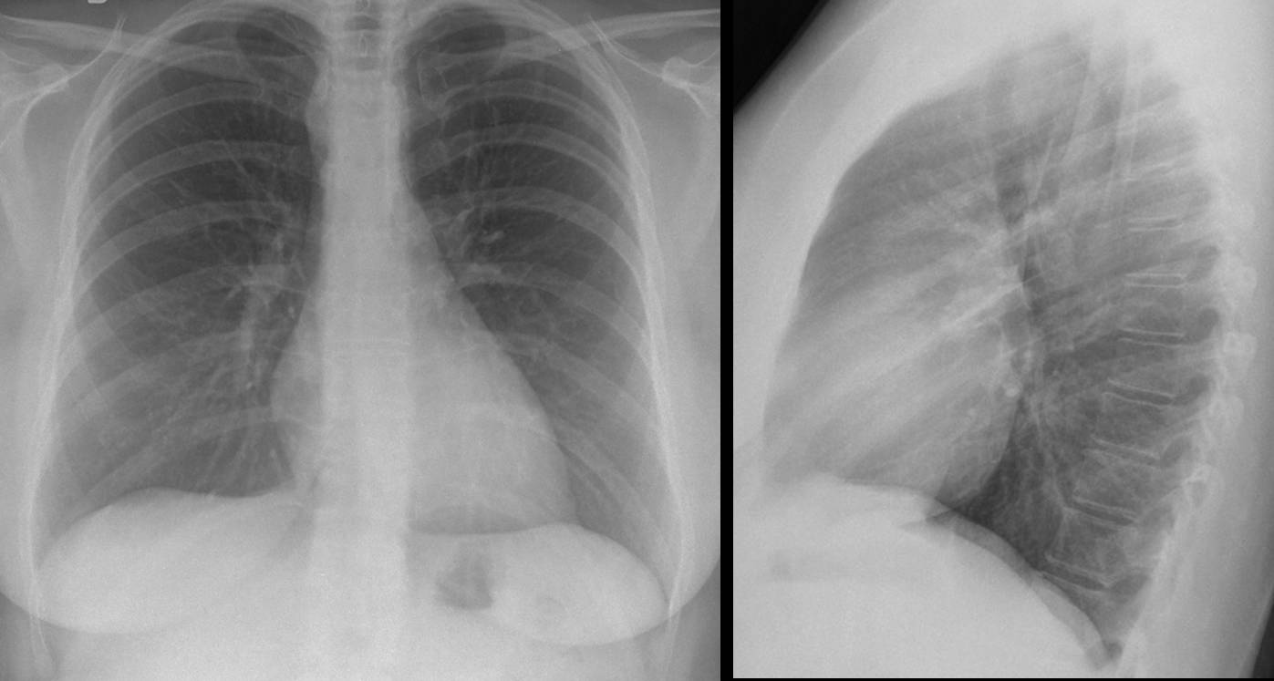
As keen followers of my career will know, the chest is my particular area of expertise, so that’s where we’re returning this week. This case is a 39 year-old woman with a cough.
1. Mediastinal lymph nodes
2. Endothoracic goiter
3. Esophageal diverticulum
4. None of the above
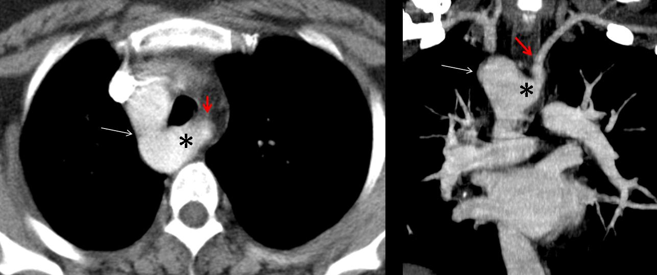
Given that the trachea is in a central position and the aortic arch cannot be seen on the left side, the most likely diagnosis is a vascular anomaly (
right aortic arch). CT confirms the right aortic arch (long arrow). The left subclavian artery (
red arrow) is seen to arise from a large Kommerell diverticulum (asterisk).
Final diagnosis: Right aortic arch with aberrant left subclavian artery
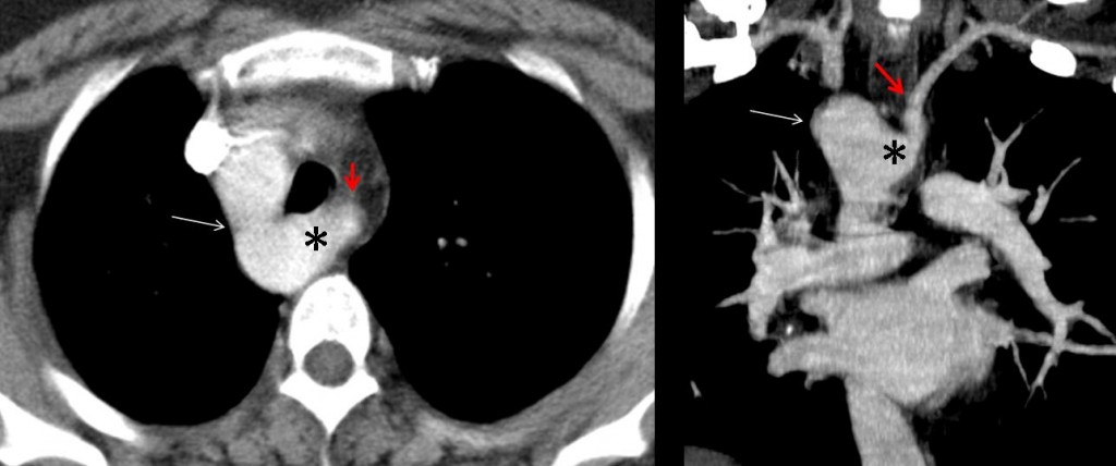
Fig. 2
Right aortic arch is one of the congenital aortic variants. Congenital aortic arch anomalies (CAAA) result from errors in the embryological development of the branchial arches, and include isolated aberrant right subclavian artery, right aortic arch, and double aortic arch. CAAA are often discovered as an incidental finding on chest plain films performed for other reasons.
Right aortic arch can be divided into two groups. Those without an aberrant right subclavian artery (mirror image) are often symptomatic because they are associated with cardiac anomalies, whereas those with an aberrant right subclavian artery only sporadically show symptoms in adults (stridor, wheezing or dysphagia) due to tracheoesophageal compression.
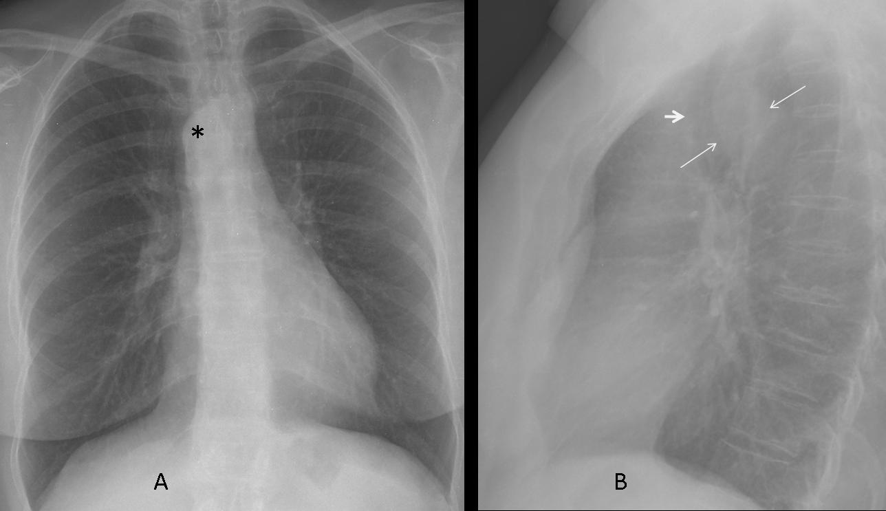
In patients with right aortic arch, the chest plain film shows the trachea in a central position or displaced to the left side, and the aortic arch cannot be identified on the left side. Sometimes a large aortic arch is seen on the right side of the trachea (asterisk in A). The lateral view (B) may show anterior displacement of the trachea (short arrow) with widening of the posterior tracheal line (long arrows).
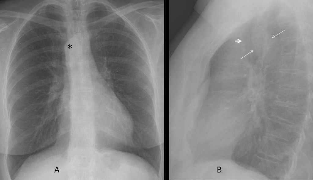
Fig. 3
CT and MRI are excellent tools to differentiate between congenital aortic arch anomalies, and are the main diagnostic techniques used in symptomatic patients:
49 year-old woman with asthma. The plain film (Fig. 4) shows the trachea in a central position and a right aortic arch (arrow).

Fig. 4
CT confirms the aortic anomaly and shows an incomplete double aortic arch (Fig. 5).
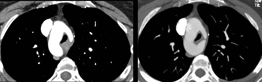
Fig. 5
This anomaly produces a vascular ring with narrowing of the trachea (Fig. 6a). The left subclavian and left common carotid arteries arise from the anterior portion of the incomplete left arch (Fig. 6b).
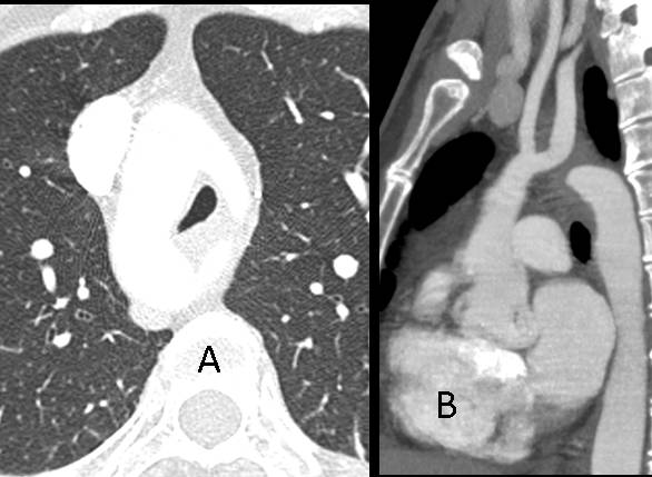
Fig. 6
The differential diagnosis includes enlargement of the thyroid gland (goiter) and paratracheal lymphadenopathy. The left aortic arch is usually visible on chest radiography and narrows down the diagnosis. When in doubt, CT can be used to differentiate between goiter, lymphadenopathy, and congenital aortic anomaly.
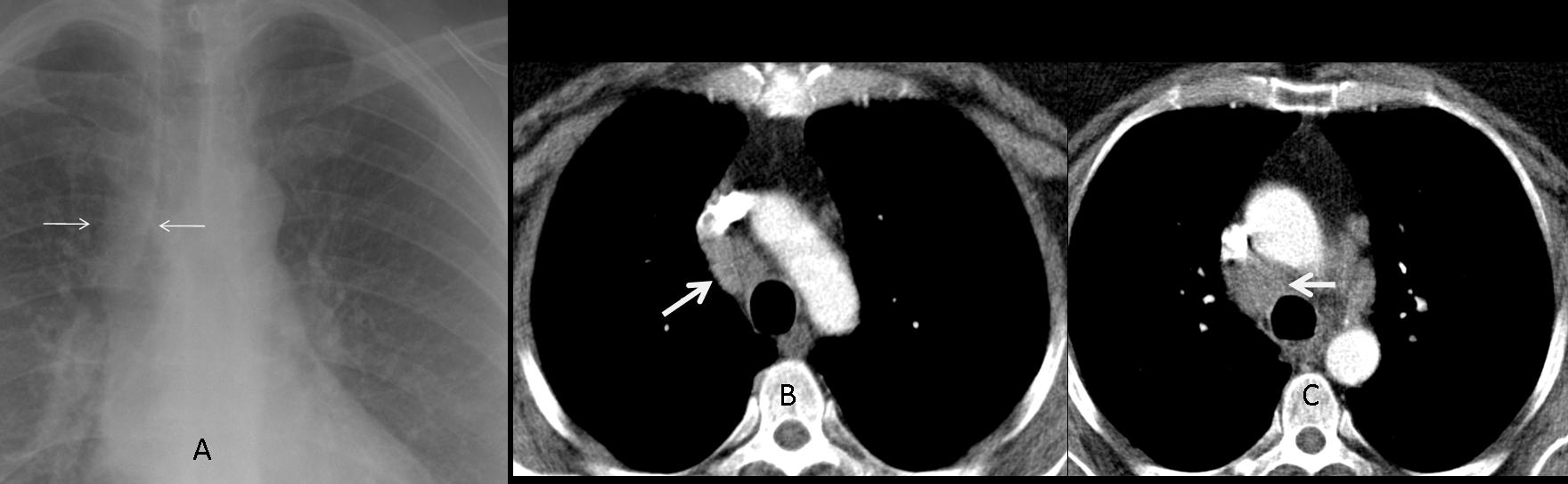
45 year-old woman with sarcoidosis. Plain film shows widening of the right paratracheal stripe (arrows, A). CT confirms lymphadenopathy in the right paratracheal region (arrows, B and C).
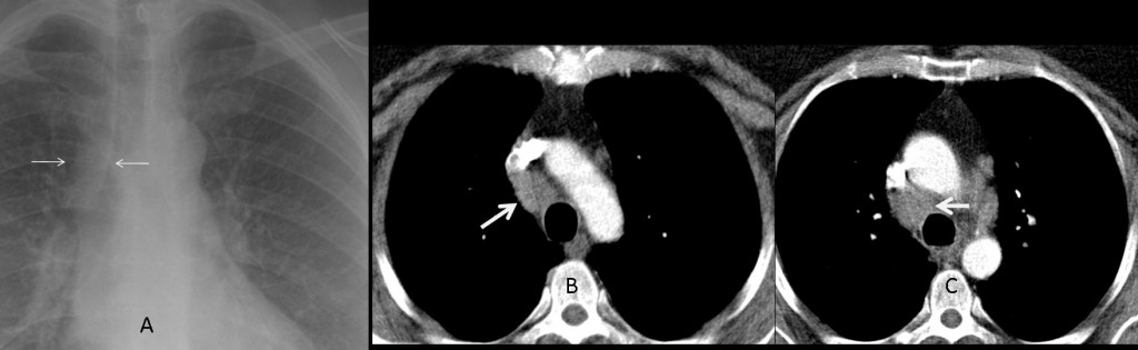
Fig. 7
52 year-old man with goiter. The plain film shows a cervical-thoracic mass (Fig. 8, asterisk) with tracheal displacement (arrows); the aortic arch is in the left position.

Fig. 8
CT confirms the presence of an enlarged thyroid (Fig. 9, arrow) with calcifications.
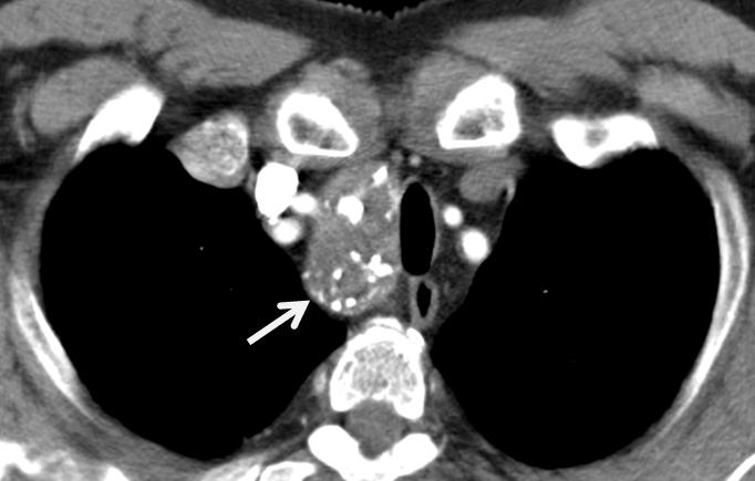
Fig. 9

Follow Dr. Pepe’s advice
Follow Dr. Pepe’s advice:
- Congenital aortic anomalies can be recognized in the chest radiograph by the position of the trachea and the aortic arch.
- CT and MRI should be used to differentiate between congenital aortic anomalies.
Ref. Embriology and imaging review of aortic arch anomalies. JTI 2012; 27:73-84
Case prepared by JM Mata