Dear friends,
This case goes to basics and it should not be difficult to interpret.
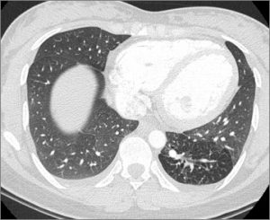
28-year-old. Incidental finding.
What finding is there and what do you think?
Check the images below!
Click here for the answer
Diagnosis: Pulmonary Sequestration
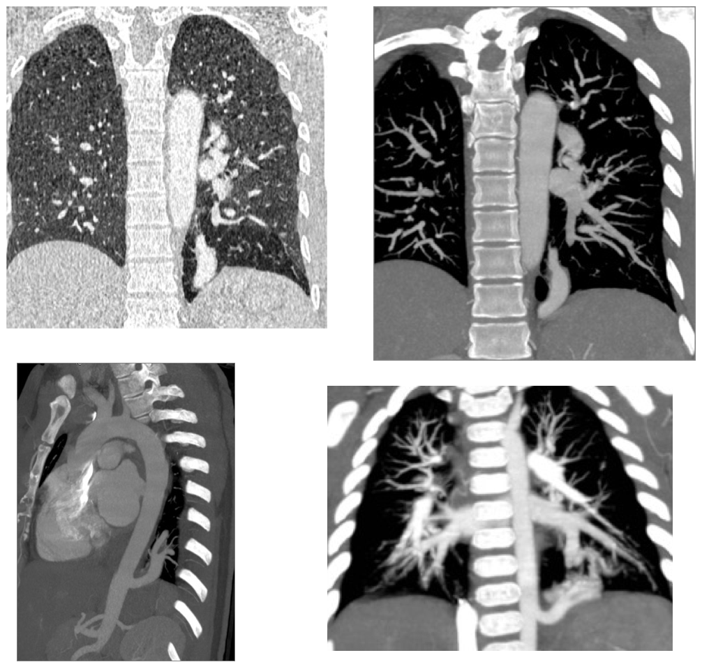
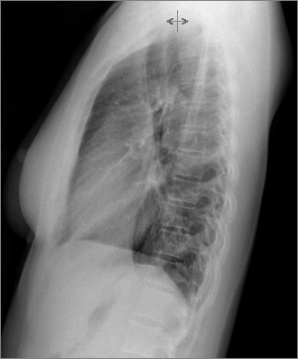
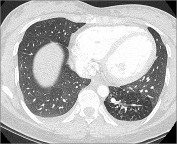
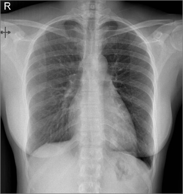
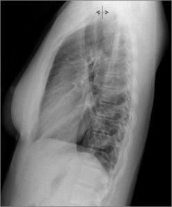
Comment: The plain films show an abnormal tubular opacity in the left lower lobe. There is some air trapping surrounding the lesion, better seen in in the CT (lung window).
The CT angiogram reveals a large vessel connected to the descending aorta.
Intralobar pulmonary sequestration shares the visceral pleura with the normal lung usually occurs in the left lower lobe. The irrigation is systemic (aorta or branches) and the venous drainage to the pulmonary veins.
Teaching point: Air trapping is common in sequestration and can help you distinguish it from AV fistulae, but remember that tubular structures with air trapping also will occur with bronchial atresia or other causes of bronchial obstruction.
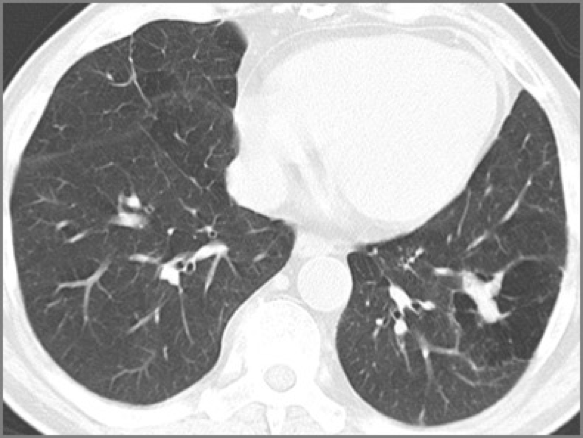
Bronchial atresia. Mucus filled bronchus simulates a vessel and air trapping.
Congratulations to those of you who mentioned the diagnosis, especially Trinity that did it immediately.