Muppet gratefully acknowledges the contribution of Dr. Jose Vizuete and Dr. Vilar, who supplied this case (Muppet also wants to point out that any complaints should be addressed to them!).
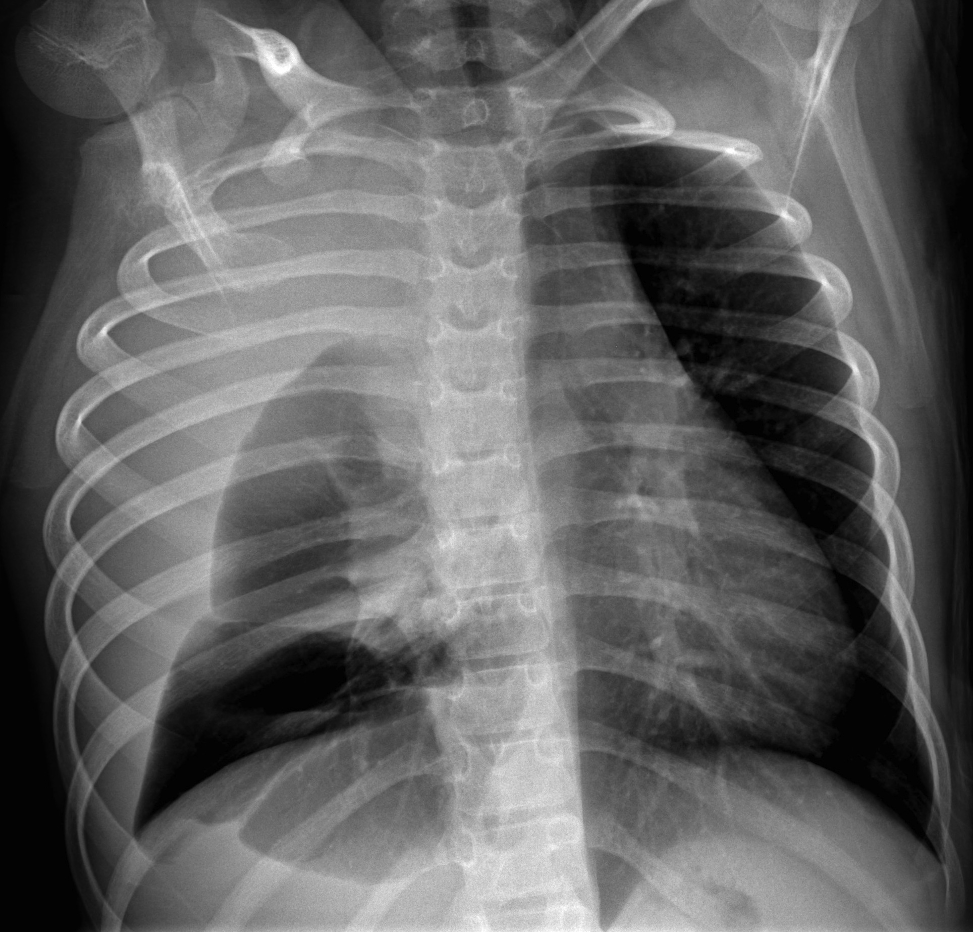
AP chest of an 11-year-old girl with dyspnea for the last week and mild fever.
1. Staphylococcal pneumonia
2. Mediastinal mass
3. Foreign body
4. None of the above
The AP chest radiograph shows partial opacification of the right hemithorax, with probable pleural effusion. The trachea and mediastinum are displaced towards the left, indicating a probable mass in the right hemithorax. The second rib is abnormal (arrows), suggesting that the mass arises from the chest wall. Findings are confirmed with CT, which shows a huge mass and the alteration of the rib (arrows).

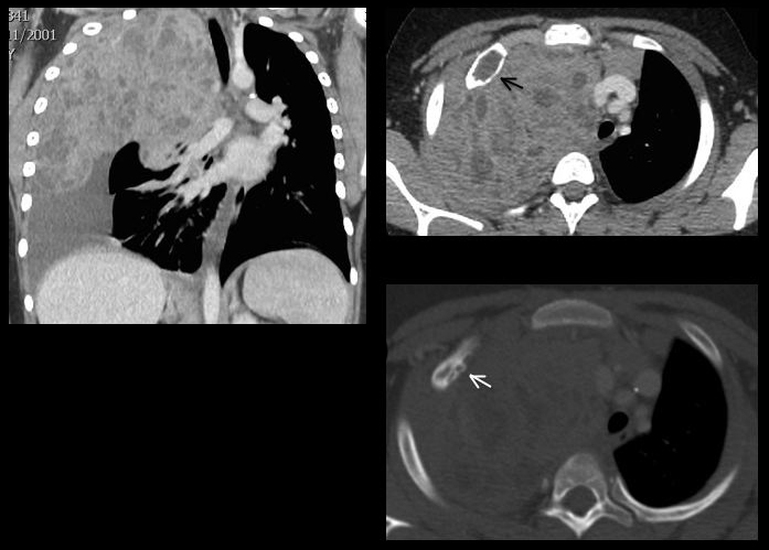
In a child, the combination of a large mass with affectation of a rib is suggestive of Ewing’s tumour or Askin tumour. Surgical diagnosis: Askin tumour.
In his (lost) youth, Muppet saw two similar cases of Askin tumour, both with large masses and not very obvious rib involvement (Fig. 3).
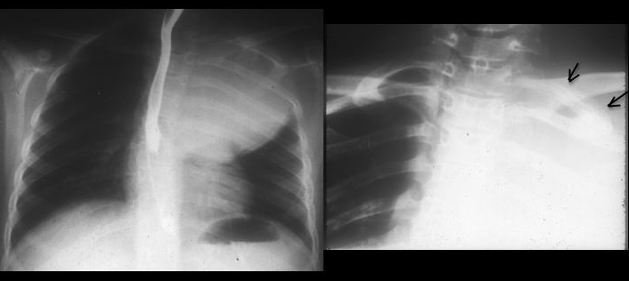
Fig 3. Askin tumour presenting as a large mass and affectation of the first left rib (arrows)
There is a similar case in the paper ‘Pediatric ribs: a spectrum of abnormalities’ (RadioGraphics Jan 2002 22:887-104). In his limited experience, Muppet suggests that if you see a child with a large tumour and limited affectation of a rib, think of Askin.
Before anybody starts complaining (case too difficult, I don´t see the abnormality, it’s easy to see the findings after looking at the CT, etc.) Muppet would like to point out that Dr. Vizuete saw the findings in the plain film and suggested the diagnosis.
Teaching point: when you see large masses that contact the chest wall, look at the ribs. They may give you a clue.