Dear Friends,
This year I plan to show only chest cases, emphasising the diagnostic approach to basic patterns in the plain film. Hopefully, this monographic approach will help you with the diploma examination.
Cases will be posted every other Monday and answers will be given on Friday.
Radiographs (below) of the first case belong to a 57-year-old man, asymptomatic. Study them carefully, leave your opinions in the comments section, and look out for the answer on Friday.
Diagnosis:
1. Thymoma
2. Teratoma
3. Mediastinal fat
4. Can’t tell
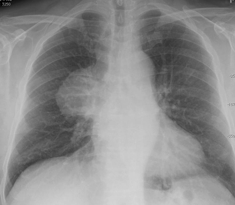
57-year-old man, asymptomatic
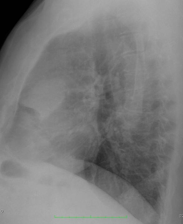
57-year-old man, asymptomatic
Click here for the answer
Findings: chest radiographs show an anterior mediastinal mass (arrows). The obvious choice is either thymoma or teratoma. However, it is important to remember that in chest plain films, all mediastinal masses have similar densities and fat cannot be distinguished from soft tissue. Therefore, the correct answer should be no. 4:
Can’t tell
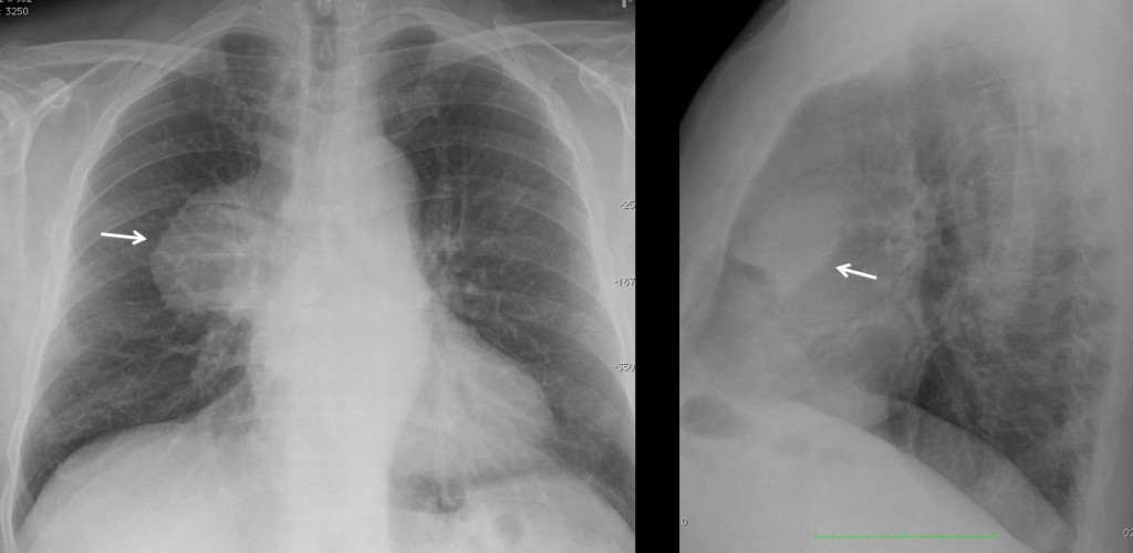
Fig. 1
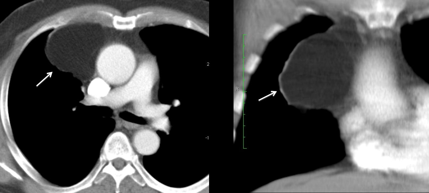
Axial and coronal CT images show a well-defined fatty mass (A,B arrows), with no internal vessels.
Final diagnosis: mediastinal fat/mediastinal lipoma
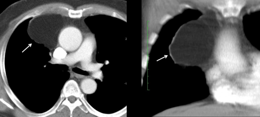
Fig. 2
This case is shown to illustrate that all mediastinal masses have similar density in plain films. To determine their radiographic density, CT must be performed.
The role of plain films in mediastinal masses is:
1. To discover them
2. To localise them in a mediastinal compartment, to narrow down the differential diagnosis
Once these steps are taken, it is imperative to do cross-sectional imaging (usually CT) to confirm the location of the mass, its radiographic density, and response to contrast enhancement.

Follow Dr. Pepe’s advice:
- The role of chest radiography in mediastinal masses is to identify and locate them.
- Mediastinal masses are characterised using enhanced cross-sectional imaging.
- The great imitators are vascular structures, mediastinal fat, and oesophageal disease.