Today we are wearing typical Basque txapelas; a gift from my good friend Dr. Estibalitz Montejo from Bilbao.
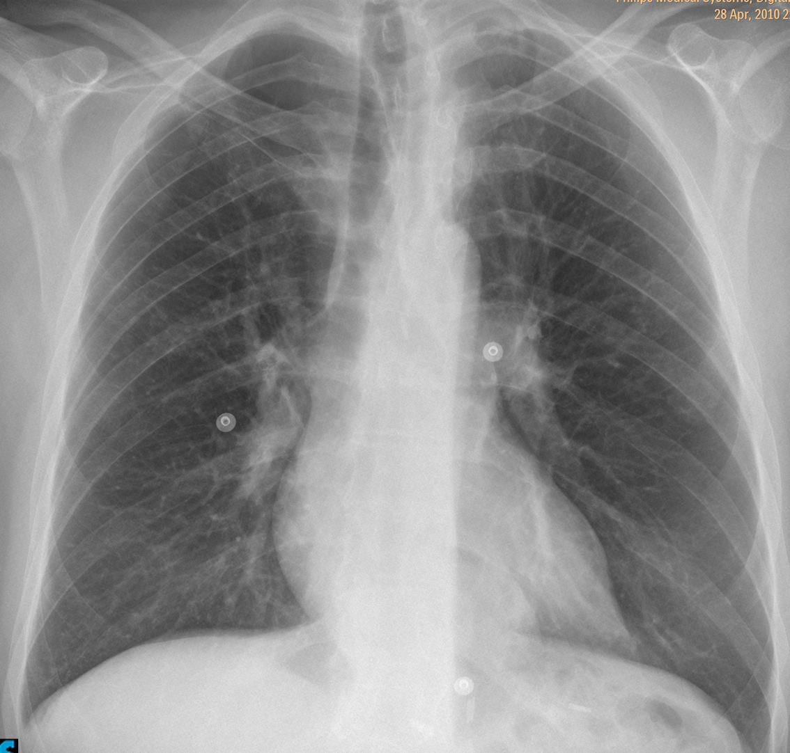
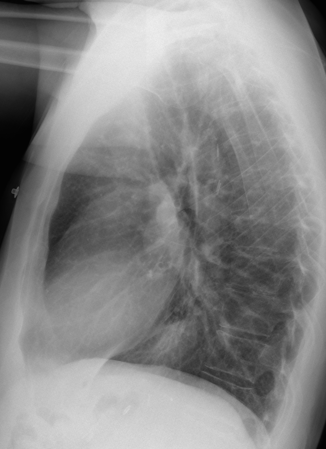
Below are two radiographs of a 45-year-old male with acute chest pain. What do you see? What would be your diagnosis?
Leave us yout thoughts in the comments section and come back on Friday for the answer.
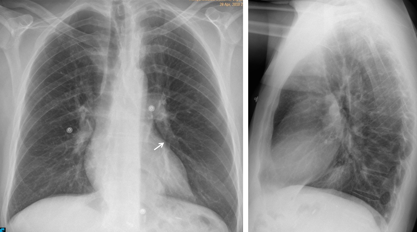
Findings: the most obvious finding is redistribution of the pulmonary vasculature, with increased size of the upper lobe vessels compared to the lower ones. This appearance has been described in mitral disease, left heart failure and emphysema of lower lobes, among others. In this particular case, the appearance of the chest does not support these diagnoses. Redistribution may also occur in lower lobe embolism, suspected in this case for the clinical symptoms and the presence of a Westermark sign in the left lower lobe artery (arrow).
CT angiogram confirms extensive pulmonary embolism. Follow-up one year later shows a normal arterial tree.
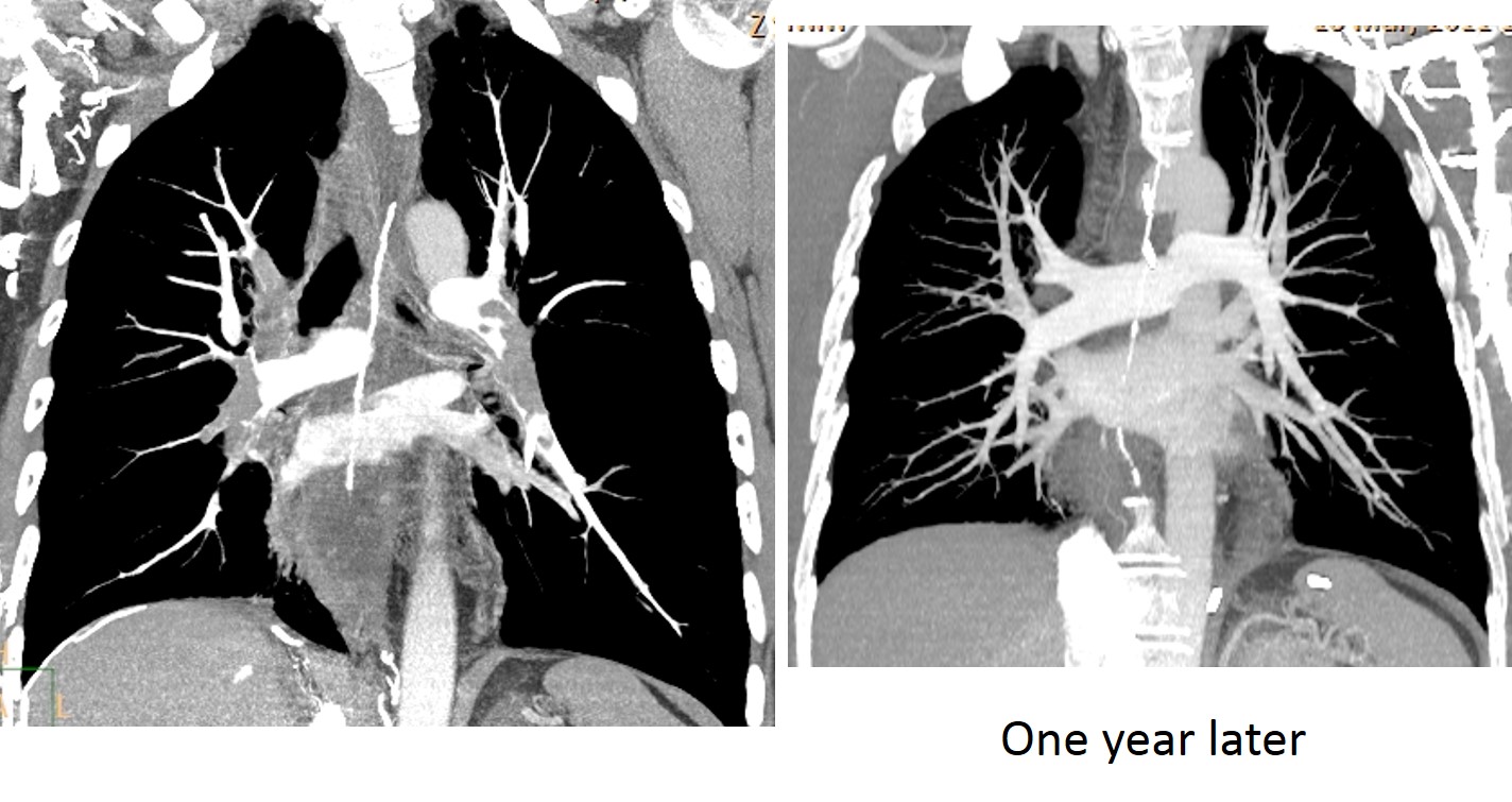
The patient had SVC thrombosis secondary to previous port-a-cath and, at the time of this episode, had right femoral vein thrombus.
Final diagnosis: acute pulmonary embolism with Westermark sign.
Congratulations to Luis Cueto, Laurens and Genchi Bari, who suggested the correct diagnosis.
Teaching point: It is important to identify vascular redistribution, because it narrows the diagnostic possibilities. After saying that, I must confess that this is my first Westermark sign!