Dear Friends,
Here is case number 8:
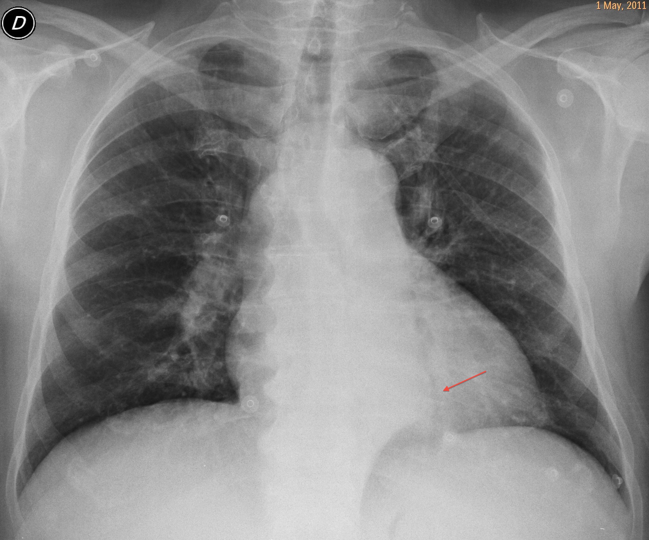
Sixty-three year old male admitted to the Emergency Room with possible pulmonary embolism. Chest radiographs show a bulge in the descending aorta. Enhanced CT from the apex of the lungs to the diaphragmatic domes confirms the embolism. A more caudal axial CT shows a rounded opacity surrounding the aorta.
Diagnosis:
1. Neurofibromatosis
2. Lymphoma
3. Cystic lymphangioma
4. None of the above
Muppet is very frustrated about the previous case. He thought you would all diagnose it easily and says that if you don’t get the new one, he will retire to a nunnery…
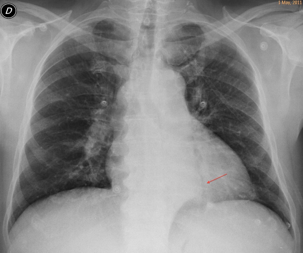
63 year old male, PA chest
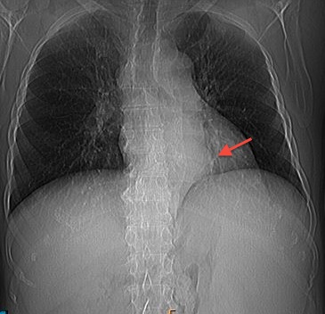
63 year old male, chest CT
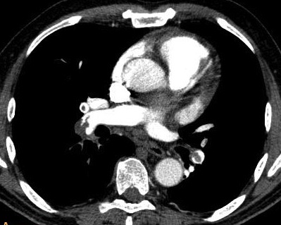
63 year old male, enhanced CT
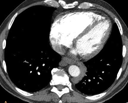
63 year old male, caudal CT
Click here for the answer to case #8
Aside from confirming the pulmonary embolism, CT reveals a rounded lesion around the aorta. The serpiginous appearance of the peri-aortic density suggests vascular structures. The prominent spleen and the enlarged azygos vein suggest portal hypertension with mediastinal varices. Diagnosis is confirmed with a delayed film, which shows enhancement of the varices (
arrows).
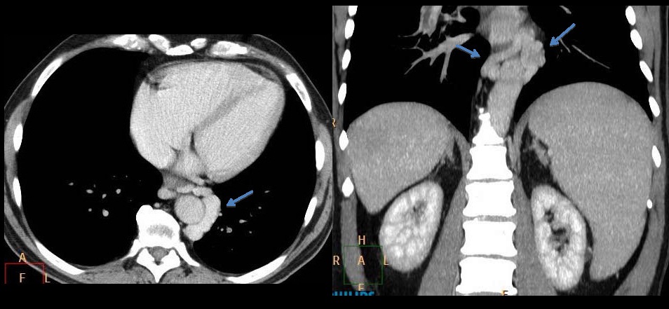
Answer Case 8
We found out later that this patient had chronic liver disease secondary to an extensive liver infarct years earlier. Mediastinal varices are not uncommon in chronic liver disease and are easily diagnosed by CT. In this particular case, because of the suspected pulmonary embolism, an early arterial phase CT was obtained, delaying the filling of the varicose veins.
Teaching point: the mediastinum is composed mainly of vascular structures. When a mediastinal abnormality is present, always rule out a vascular origin (arterial or venous).
Muppet wishes you the best for the coming year!