Dear friends,
Presenting a case chosen by the Muppet (beware: he is devious!).
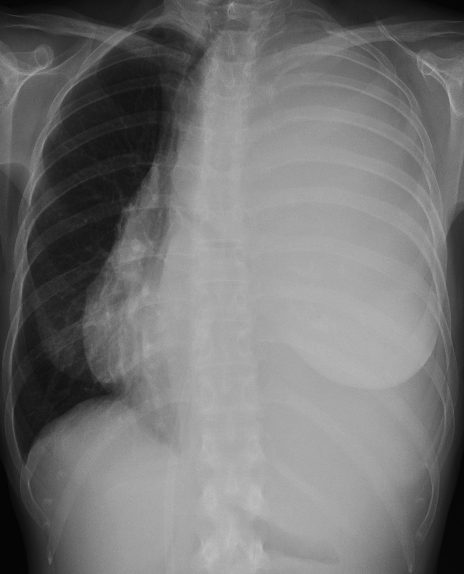
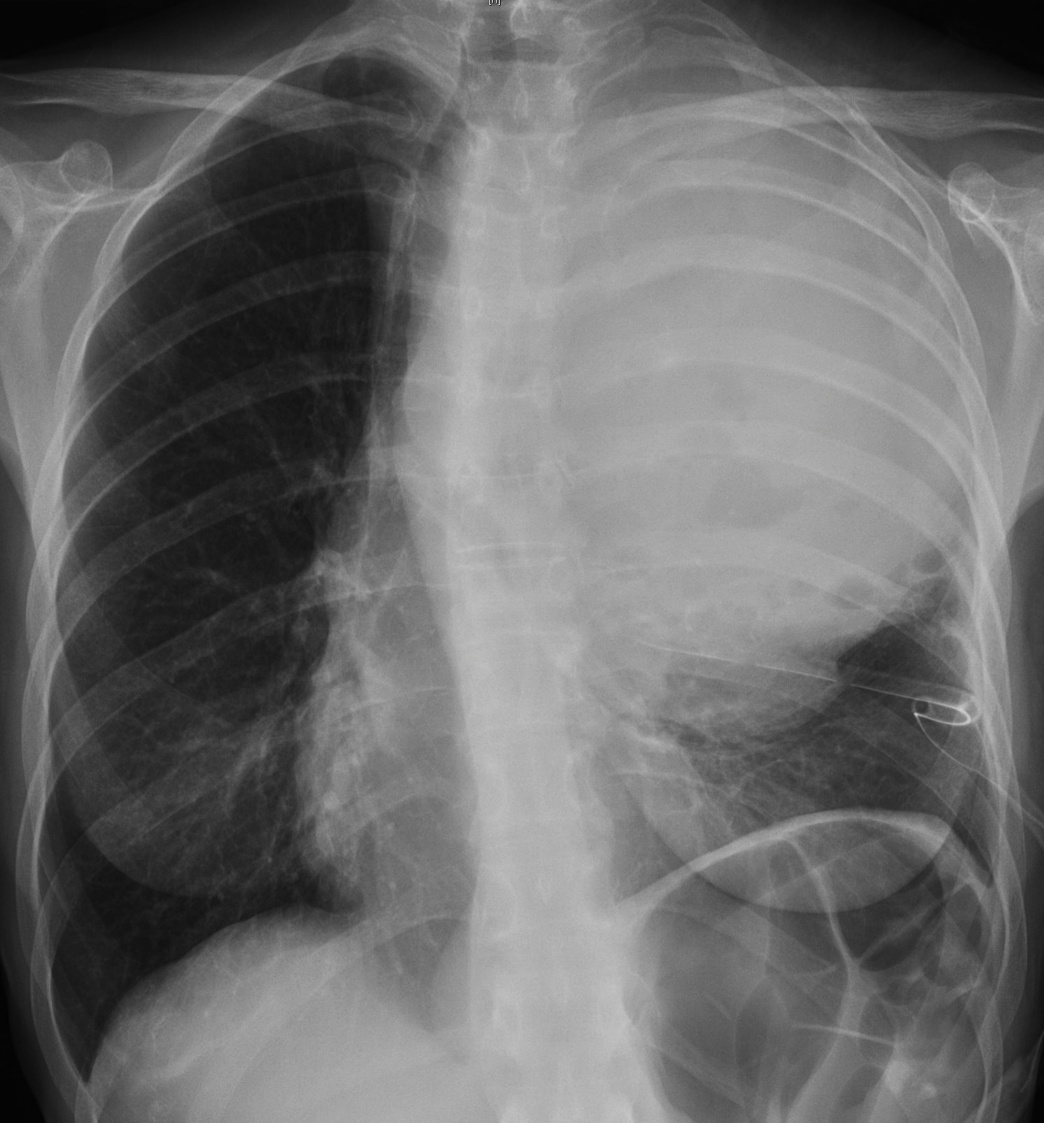
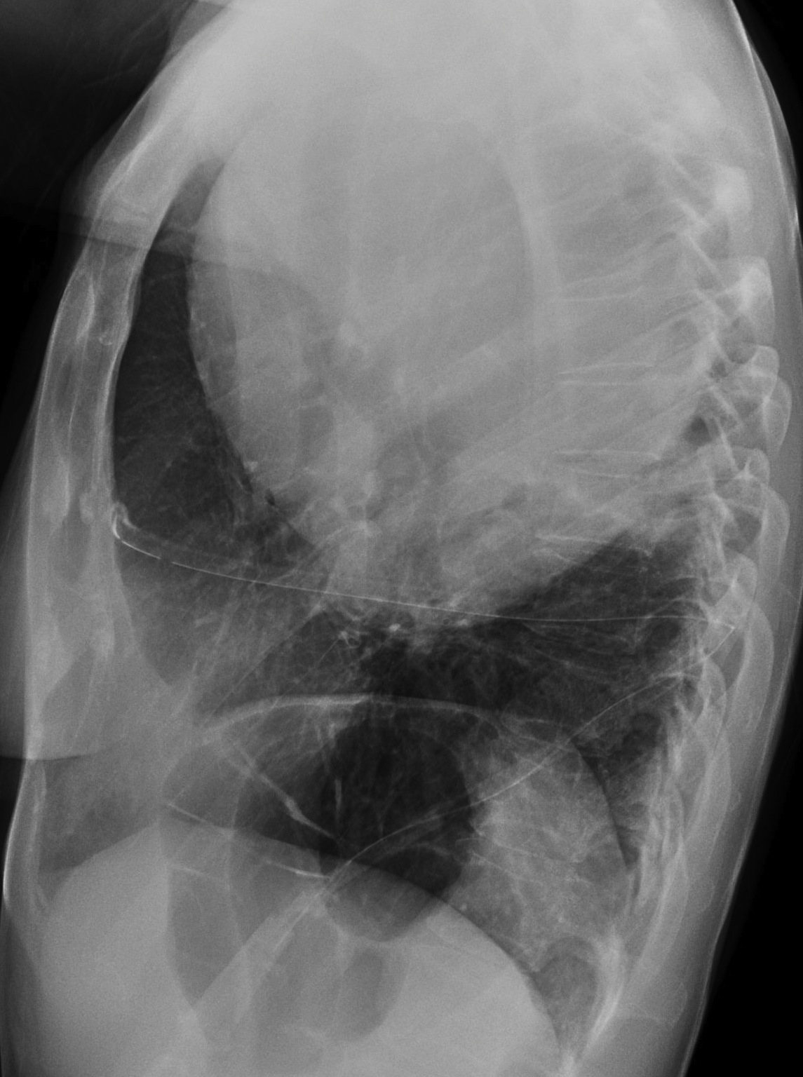
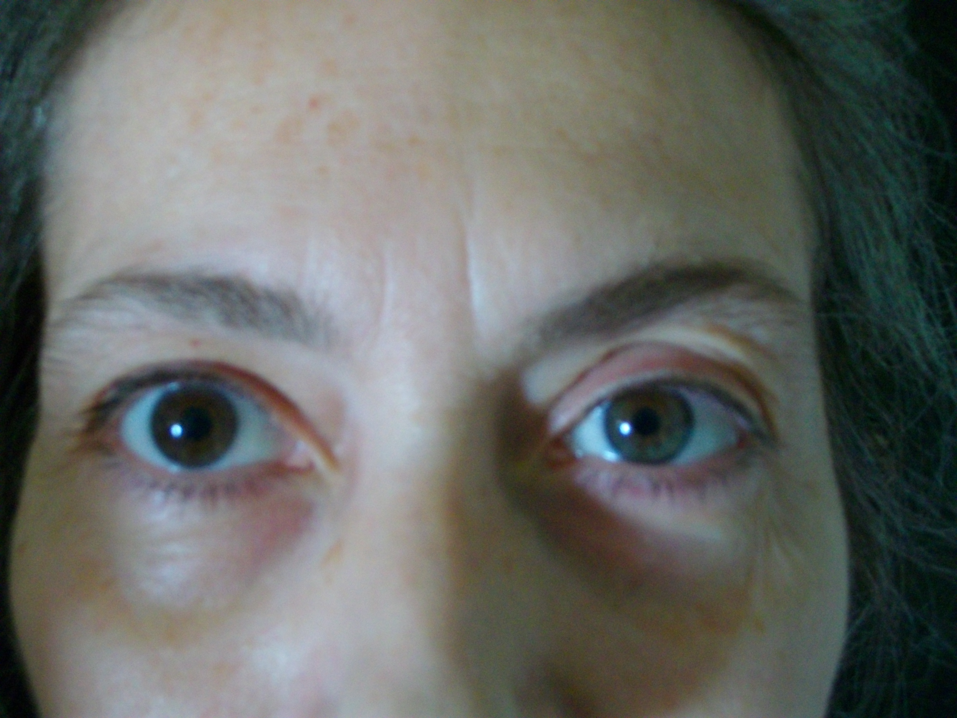
Radiographs and face picture of a 52-year-old woman admitted to the emergency room with dyspnea. Radiographs before and after thoracentesis.
Chest radiograph seven years earlier (not available) reported as possible thymoma. No treatment since.
Diagnosis:
1. Thymoma
2. Schwannoma
3. Fibrous mesothelioma
4. Hydatid cyst
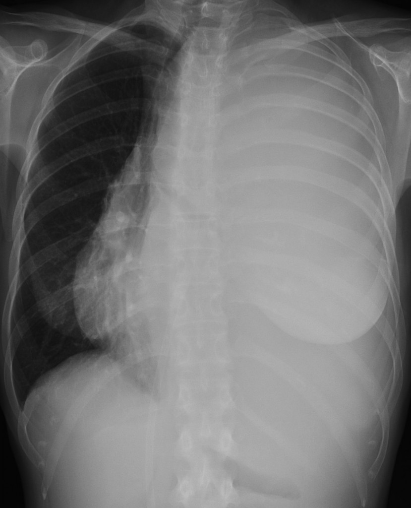
chest, 52 year old female (PA)
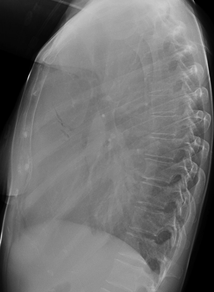
chest, 52 year old female (lateral)
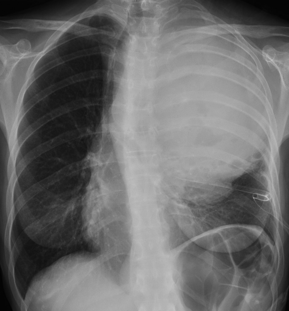
chest after thoracocentesis, 52 year old female (PA)
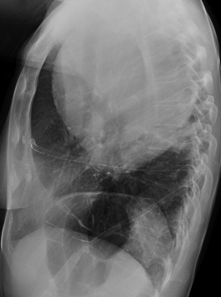
chest after thoracocentesis, 52 year old female (lateral)
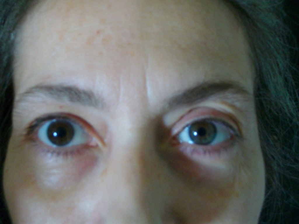
photo
Click here for the answer to case #7
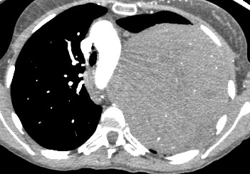
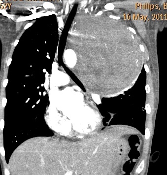
The initial radiographs show massive pleural effusion. After thoracentesis a huge mass is seen. Enhanced CT confirms the presence of the mass, without any specific characteristics. There is no bone erosion.
Diagnosis is suggested by the heterochromia iridis, a finding associated with cervical neurogenic tumours, which may affect the sympathetic system, responsible for eye colour. Operative diagnosis: schwannoma.
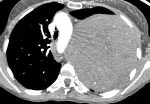
Axial CT
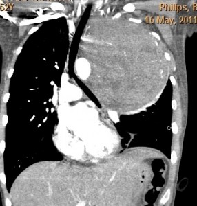
Coronal CT
We saw a similar case about fifteen years ago. We didn’t know what it was, but searched the web and came out with the correct answer. When we found this recent case, the diagnosis was evident. Muppet is disappointed that you didn’t notice the heterochromia iridis and promises not to do it again… until the next time (told you he is devious and likes to play dirty!)
Teaching point: the apex of the lung is a small space. In the presence of a large apical mass it is very difficult to determine its origin (pleural, pulmonary or mediastinal). Sometimes other findings (heterochromia in this case) point to the correct diagnosis.