Dear friends,
The Muppet is getting bored with me and has decided to invite a new player every month. Our first guest is José Vilar, a good friend and excellent teacher who has chosen the following case:
“41 year old male with thalassemia minor who had a CT as a routine follow-up after an episode of pulmonary embolism three months previously.”
Diagnosis:
1. Pleural plaques
2. Fibrous mesothelioma
3. Scarring post-infarction
4. None of the above
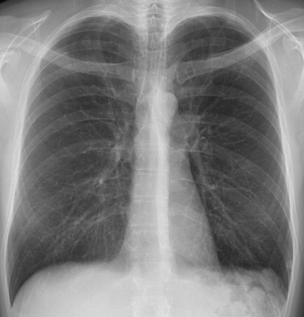
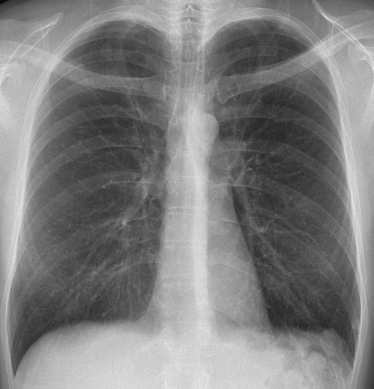
41 year old male, PA chest
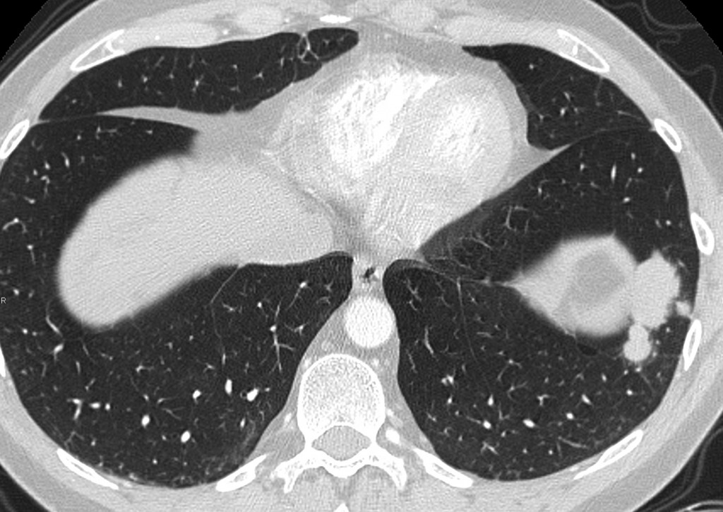
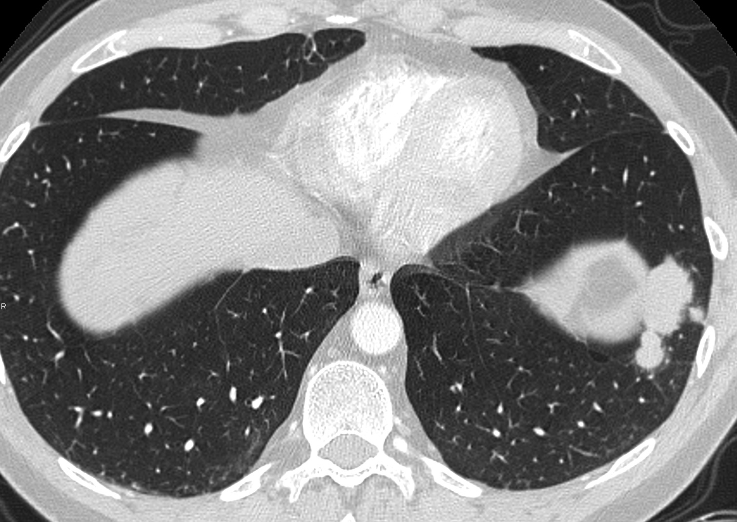
41 year old male, axial CT
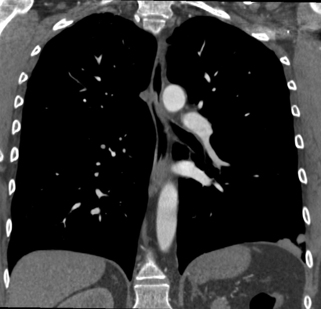
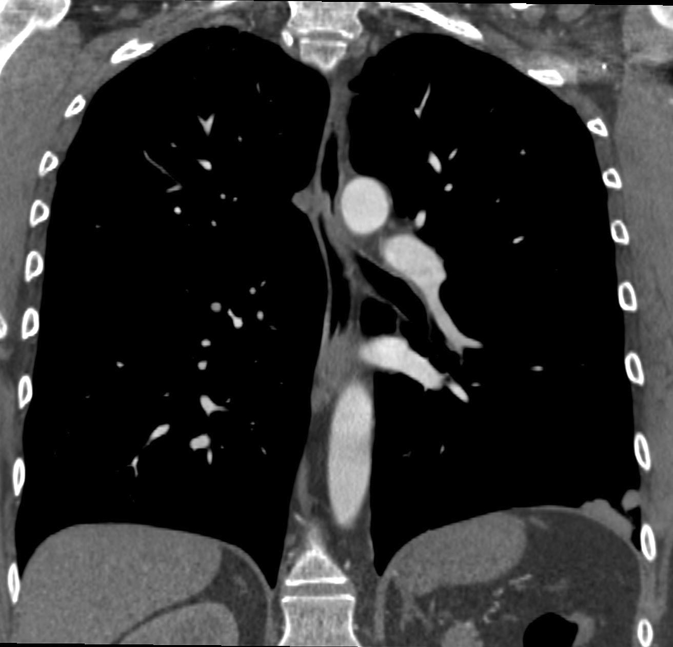
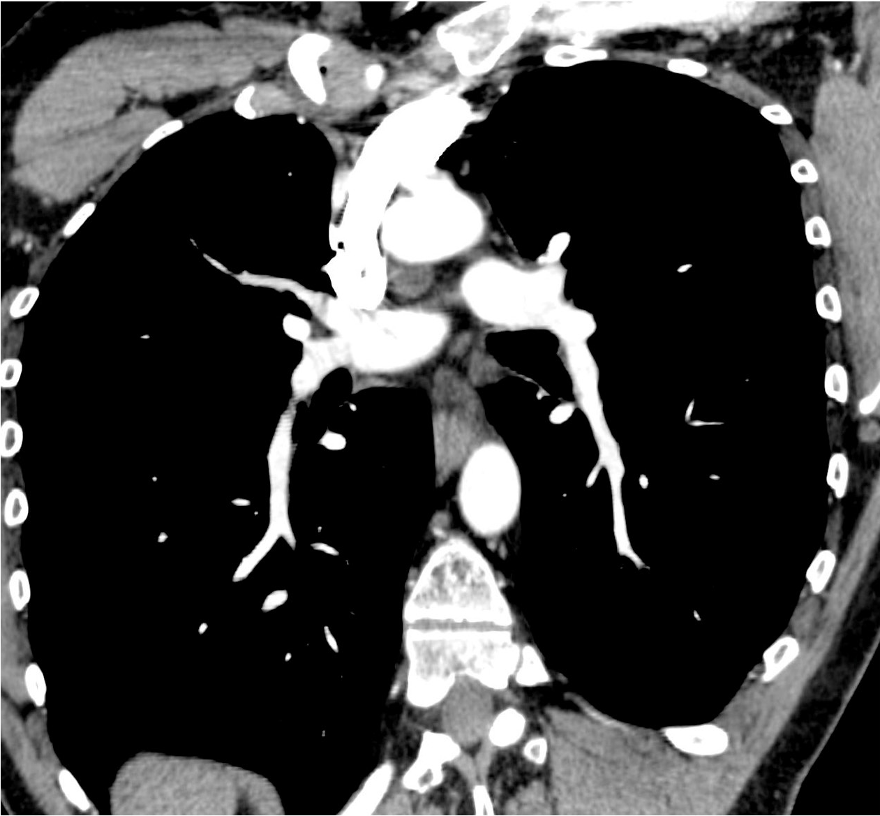
41 year old male, enhanced coronal CT
Click here for the answer to case #10
Dr. Vilar says:
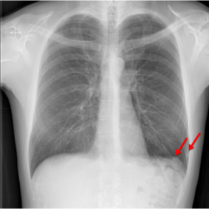
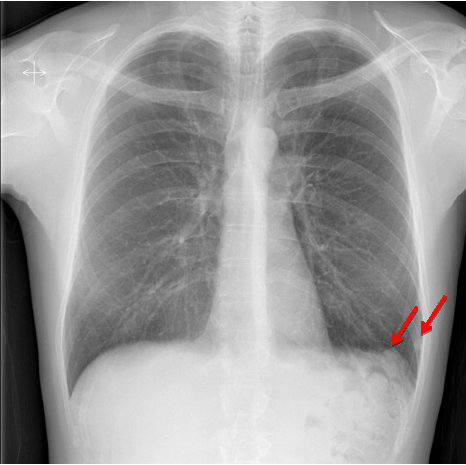
The PA radiograph shows some nodular densities near the left cardiophrenic angle.
The spleen shadow is not identified.
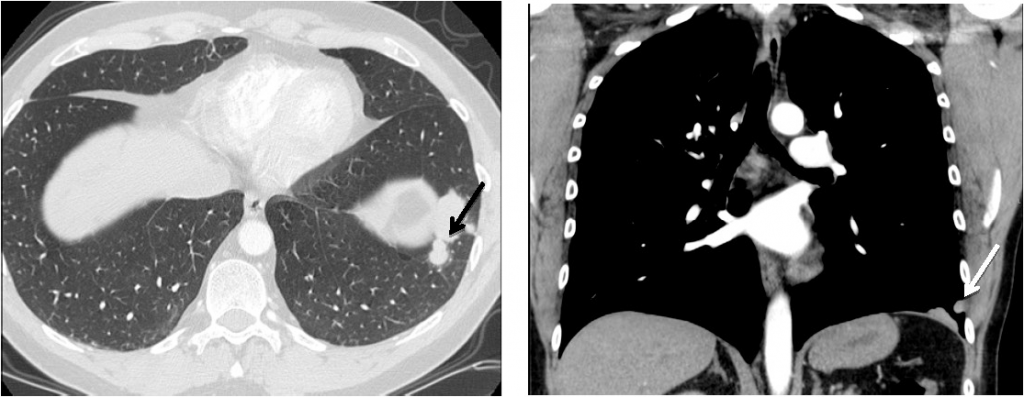
Axial and coronal CT views in an early angiographic phase show the presence of nodular lesions in the surface of the pleura.
The coronal view shows an absence of the spleen in the left infradiaphragmatic region.
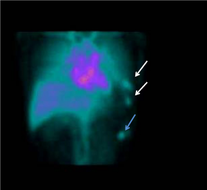
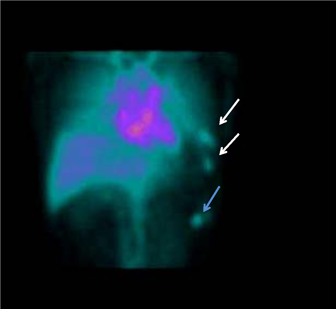
The nuclear Medicine study performed with the patient´s erythrocytes marked with Tc 99m shows the typical uptake in the the splenic nodules similar to that of the liver.
Note the presence of splenosis above ( white arrows) and below the diaphragm (blue arrow)
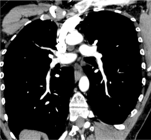
The CTA did not demonstrate any residual pulmonary embolism.
A further investigation showed that the patient had had a left nephrectomy and splenectomy after an accident several years previously.
Final diagnosis: Pleural splenosis.
Teaching point: Pleural nodules in the left side: splenosis is a good choice if you don´t see the spleen.