Dear friends,
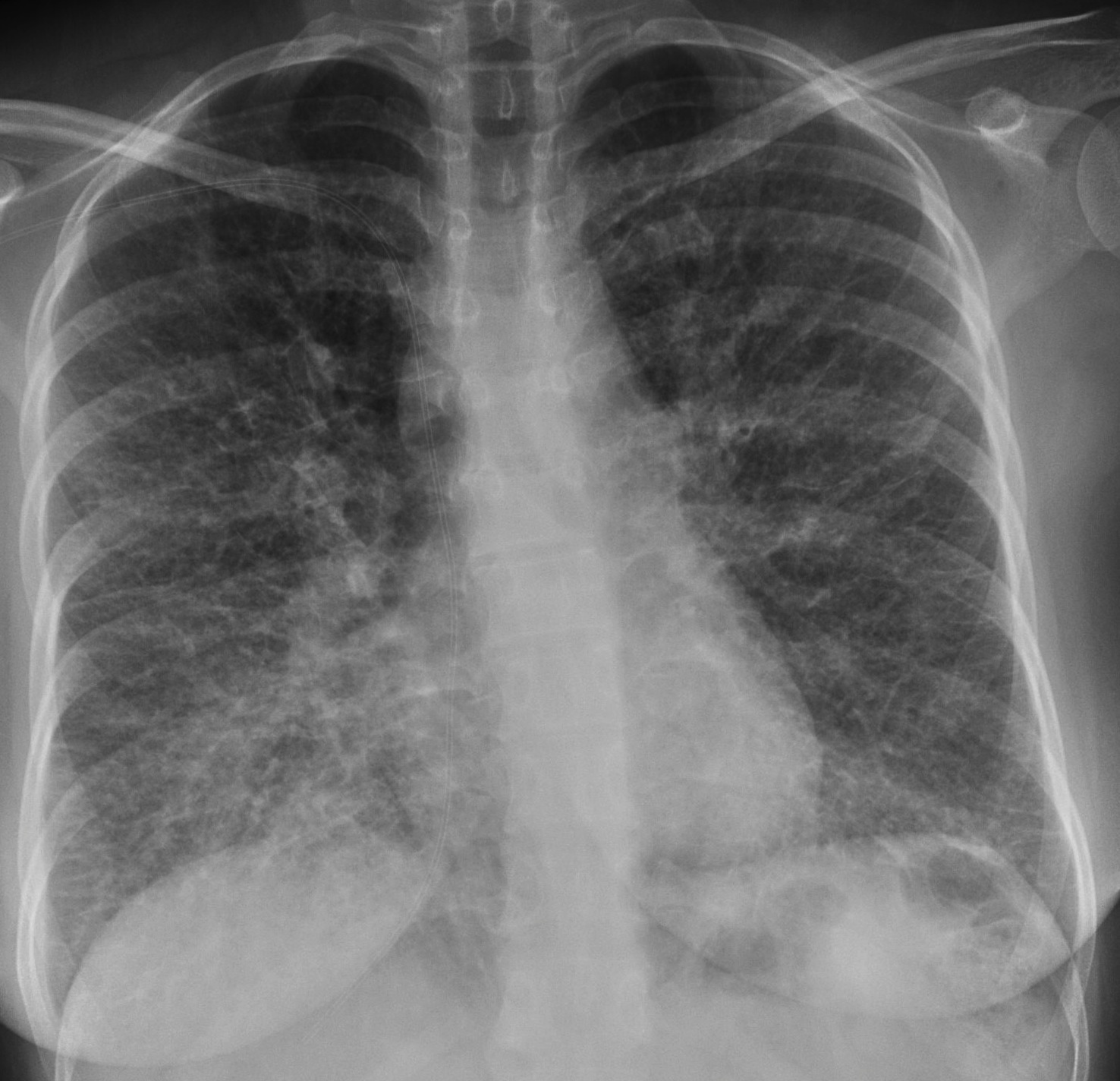
Muppet saw this case three weeks ago. Clinical history was: 36-year-old woman hairdresser, pre-op chest.
Diagnosis:
1. Sarcoidosis
2. Extrinsic allergic pneumonitis
3. Lymphangitic carcinomatosis
4. Histiocytosis X
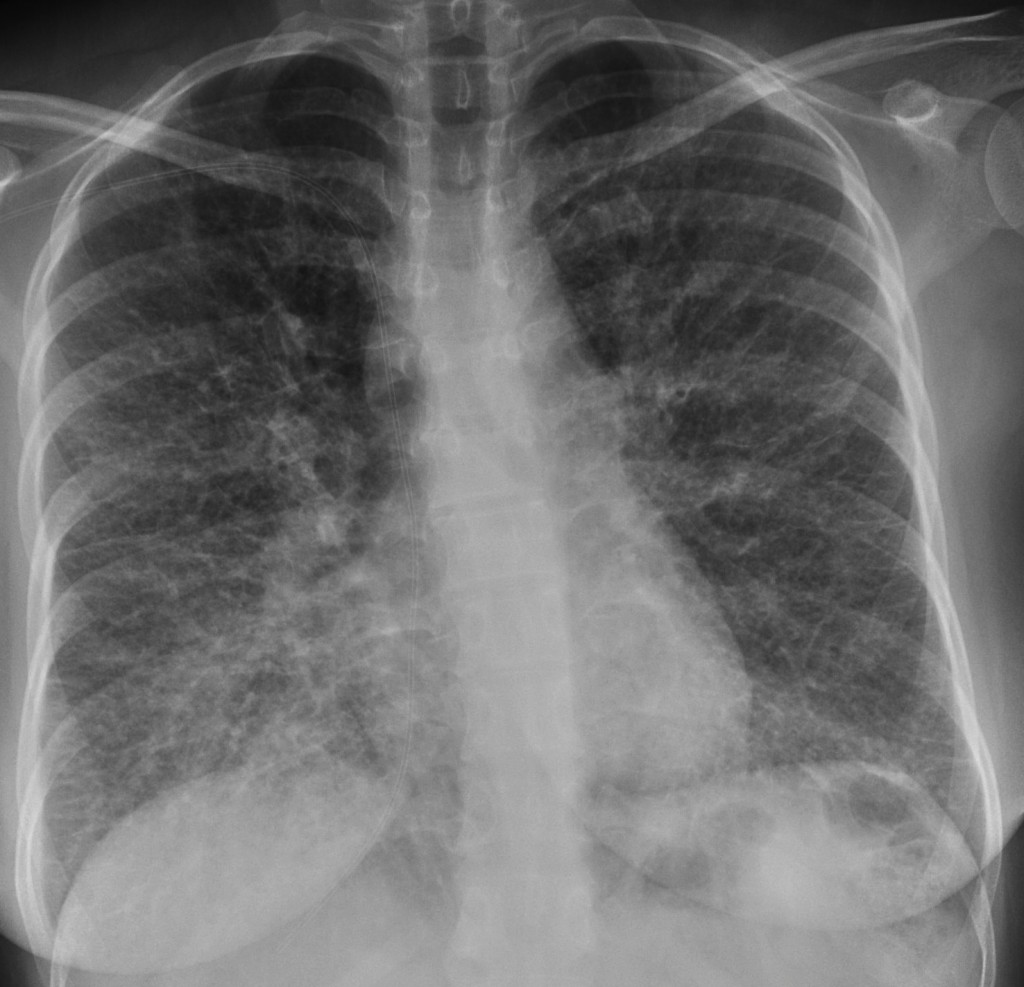
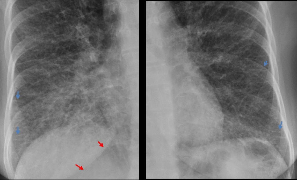
36 year old woman, PA chest
Click here for the answer to case #11
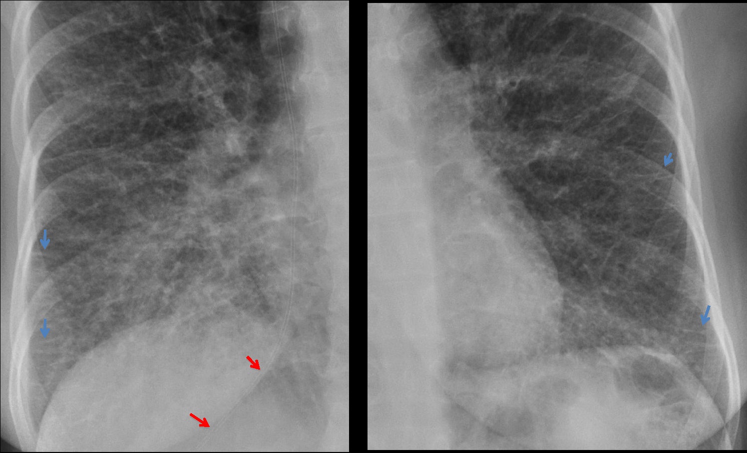
This is one of the few cases of diffuse infiltration of both lungs in which the pattern is recognisable. PA chest show the most beautiful Kerley’s lines you will ever see (arrows) identifying a pattern of diffuse lymphangitis.
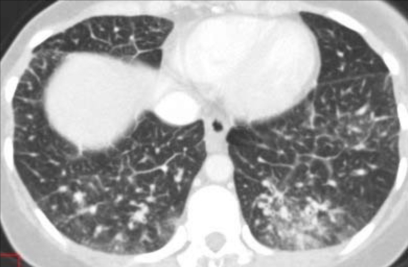
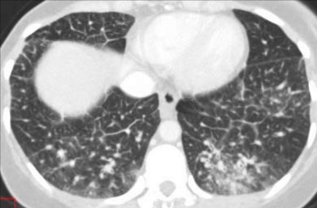
Patient had a carcinoma of the stomach diagnosed at gastroscopy. The surgeon was not aware of the lymphangitis, which was discovered in the pre-op chest radiograph. Abdominal CT for staging confirmed the lymphangitic spread of the primary tumour.
Incidentally, the venous line ends in the suprahepatic vein. I imagine you all noticed that.
Congratulations to those who made the diagnosis. Muppet hopes that the others will not forget the lesson.
Teaching point: recognising widespread Kerley’s lines in the chest film narrows down the differential diagnosis practically to two entities: cardiac failure and lymphangitic carcinomatosis.