1. Radiation pneumonitis
2. Tuberculosis
3. Carcinoma of the lung
4. Pulmonary hypertension
Answer to case # 2
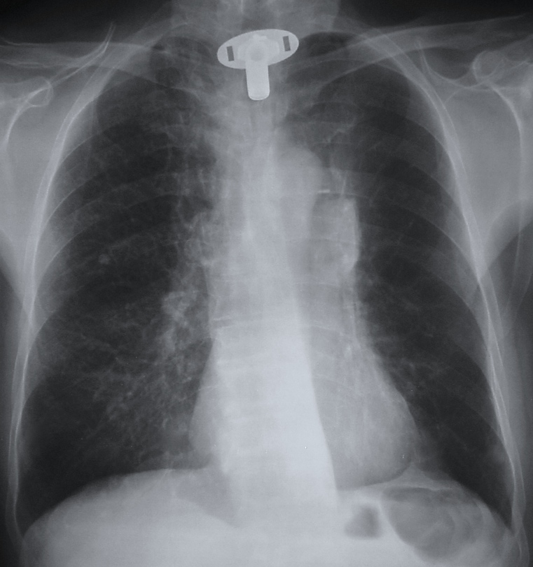
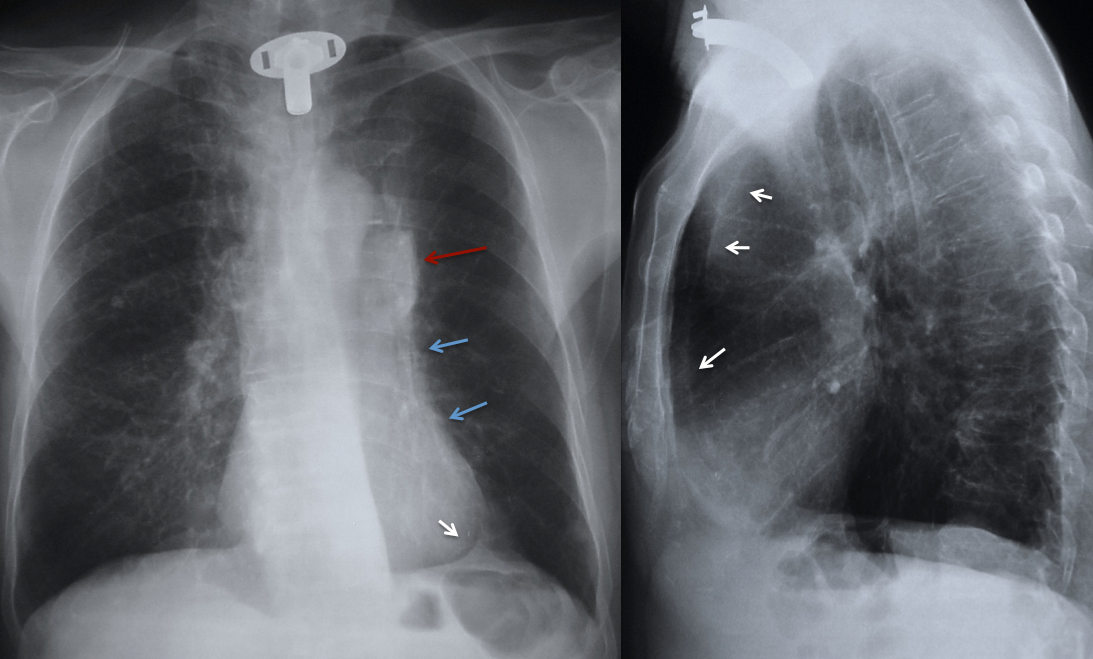
The first impression when looking at the PA chest radiograph is a prominent left hilum (red arrow). If we continue to look (we have learned to fight satisfaction of search/sex, muppet dixit), we discover that the left heart border is indistinct (blue arrows), whereas the aortic knob is well defined (luftsichel sign) and there is tenting of the left hemidiaphragm (white arrow) also called the juxtaphrenic peak sign.
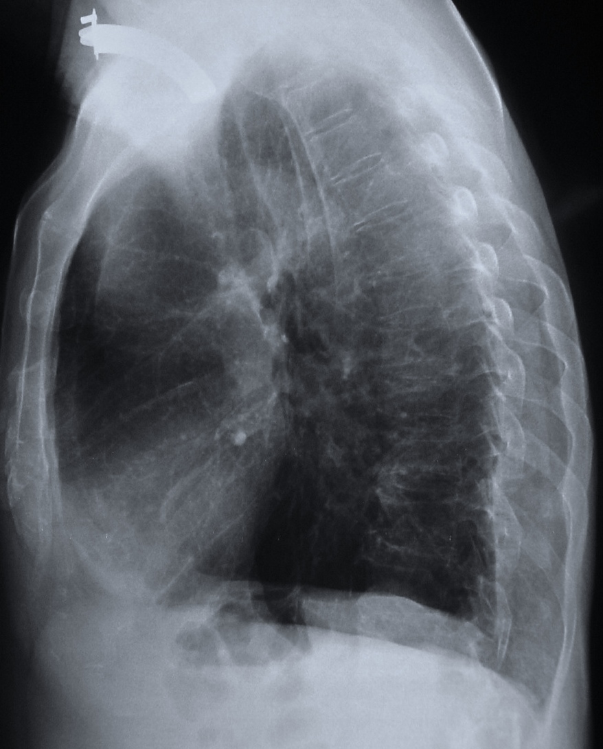
All these are typical signs of left upper lobe collapse. Diagnosis is confirmed on the lateral view, which shows a thin line behind the sternum (arrows), representing the left major fissure displaced anteriorly due to the marked loss of volume of the left upper lobe.
Once the collapse is recognised, carcinoma of the lung is by far the most likely diagnosis, since the majority of collapses of the left upper lobe are due to carcinomas.
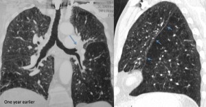
An interesting fact is that this patient had a CT one year earlier, read as normal. Review of the CT shows the endobronchial tumour (arrow) and moderate loss of volume of left upper lobe in the sagital reconstruction (arrows), both overlooked at that time.
Teaching point: important to know (and recognise, says the muppet) the signs of left upper lobe collapse because the great majority of them are due to endobronchial carcinoma
 Clinical history: asymptomatic 75 year-old man, operated on ten years ago for carcinoma of the larynx. Current chest radiographs were obtained during the yearly routine control.