Dear friends,
Here is an easy case to facilitate the transition into the new year.
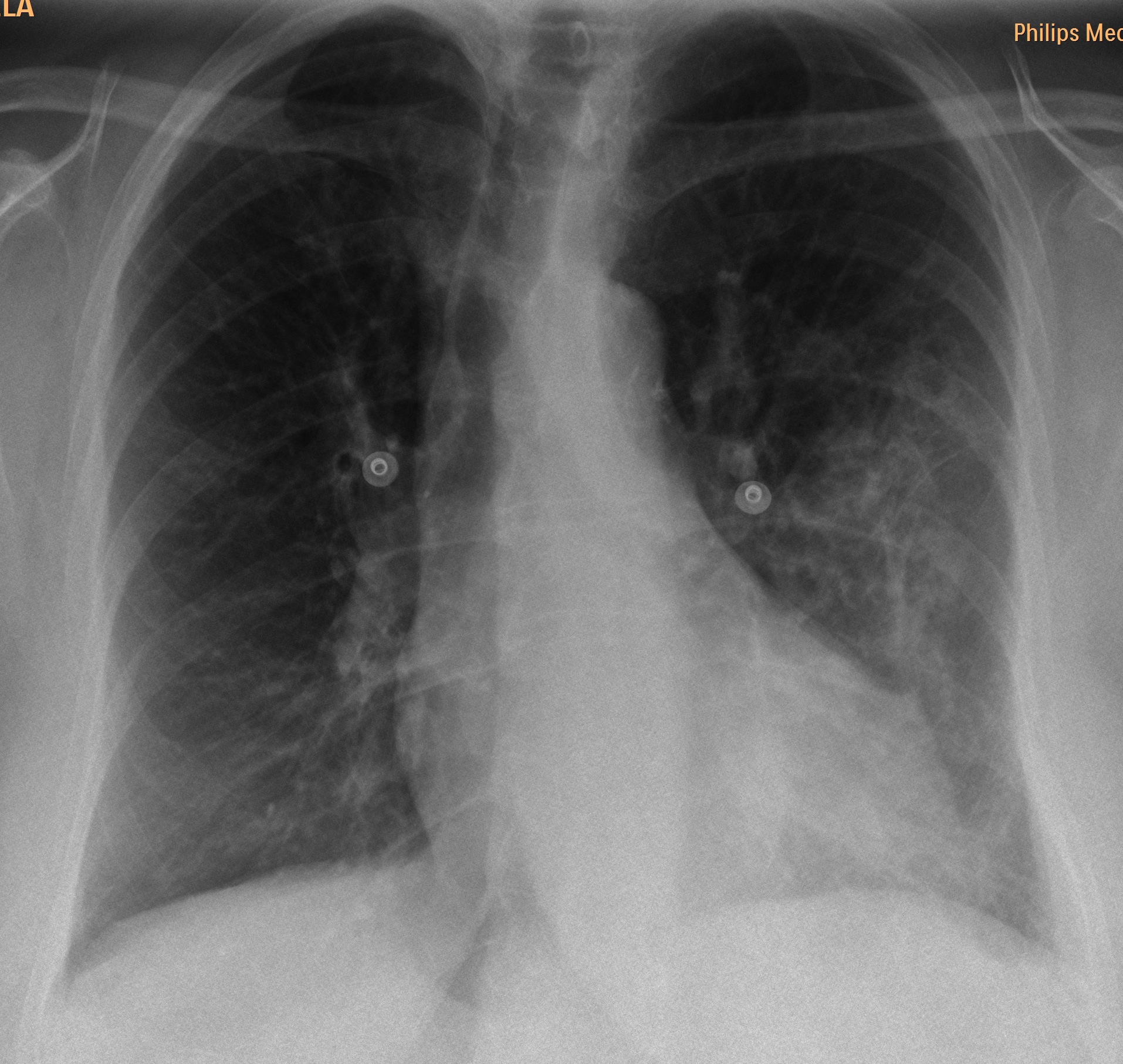
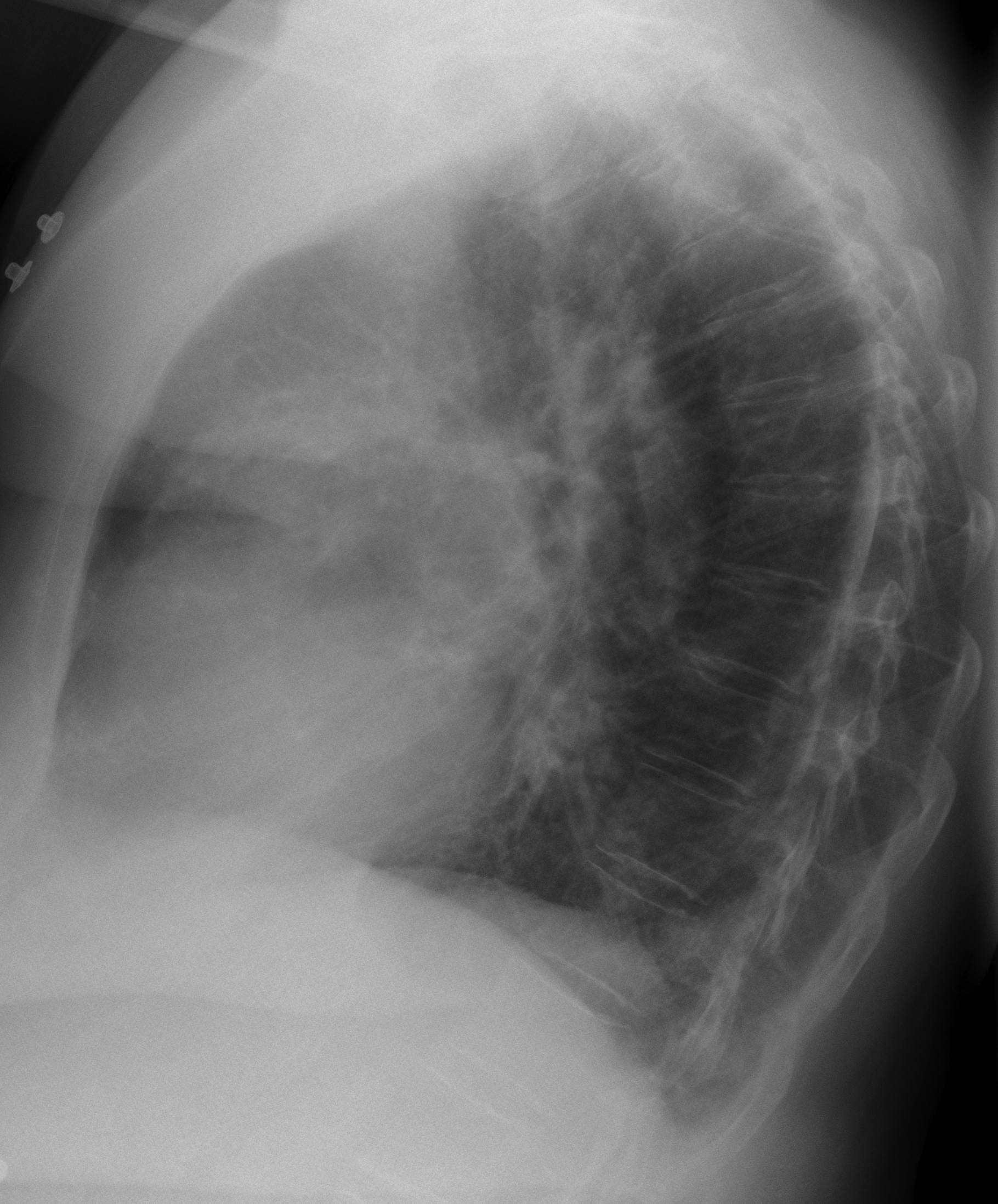
Sixty-seven year old lady with dyspnea for about a month. Previous history of bronchiectasis.
Diagnosis:
1. Infected bronchiectasis
2. Pulmonary embolism
3. Bonchioloalveolar carcinoma
4. Pleural disease
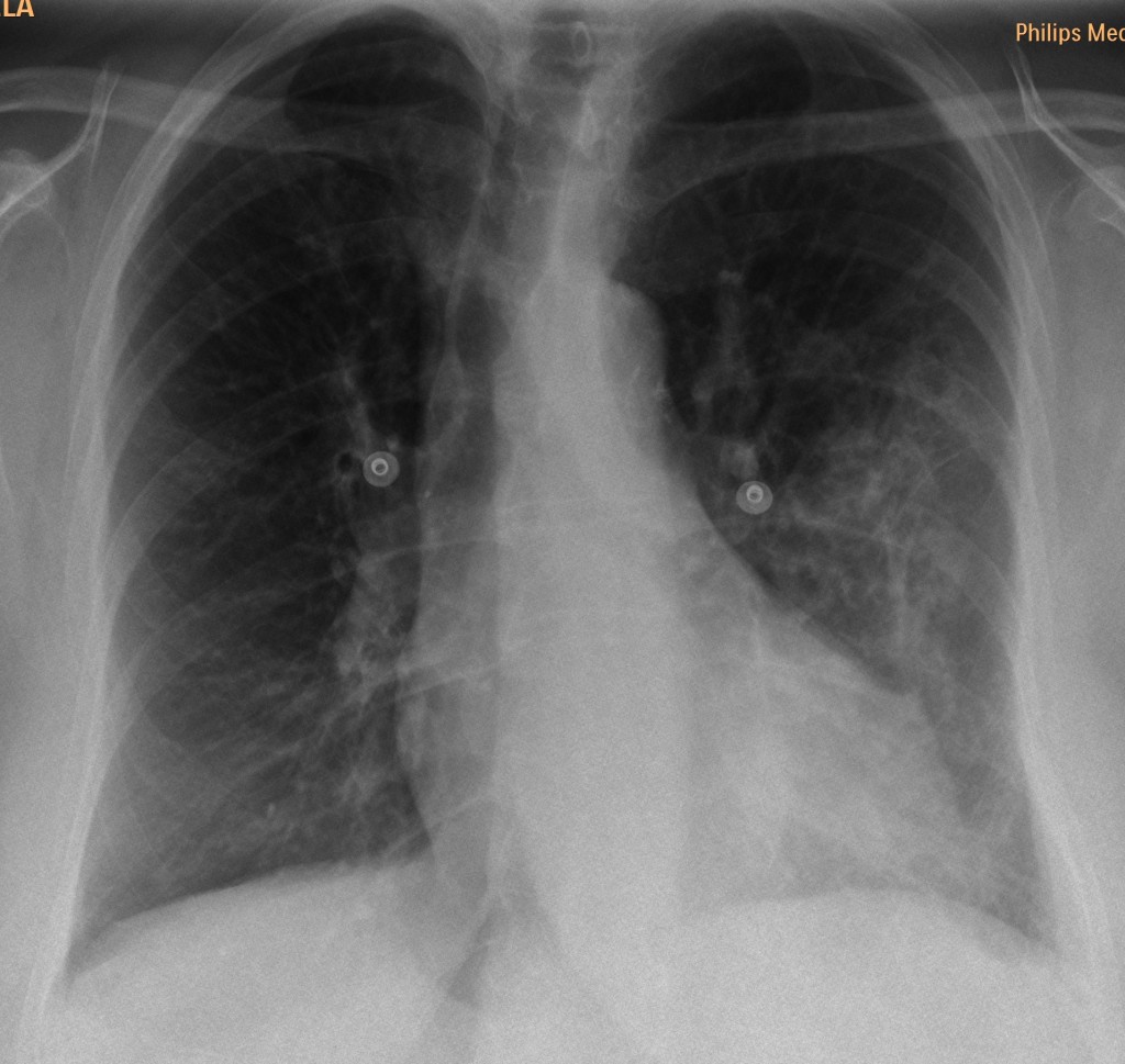
67 year old female, PA chest
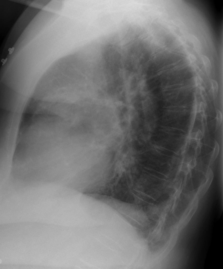
67 year old female, lateral chest
Click here for the answer to case #9
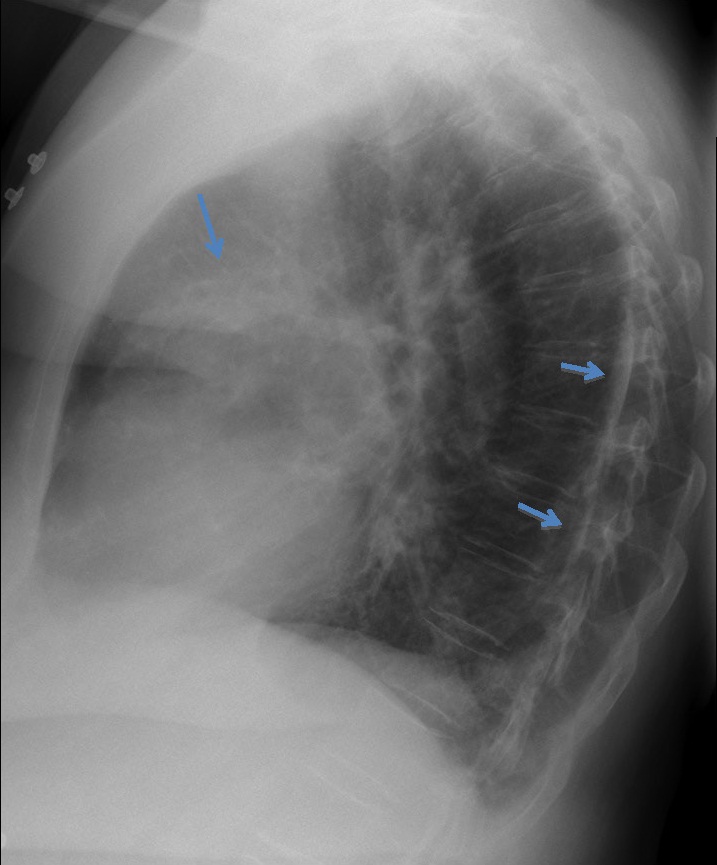
Of the four options offered, the first three cannot be diagnosed with confidence. However, obvious pleural calcification is visible on the lateral view, (arrows), which makes option 4 (pleural disease) the most likely diagnosis. This is a good example of calcified pleura seen on face on the PA view, simulating lung disease. CT confirms the diagnosis (arrows). Incidentally, no bronchiectasis was seen, which makes us wonder about the acumen of our clinical colleagues.
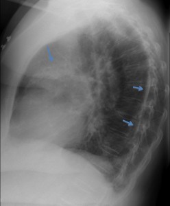
Answer case 9-1
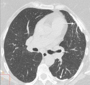
Answer case 9-2
Congratulations to all of you who made the diagnosis. It made Muppet very happy!
Teaching point: 1. Calcified pleural disease may have a bizarre appearance, which may be confused with lung disease unless one recognises it.
2. Always look at the lateral view. You never know when it’s going to help!