Welcome to Case #6. Let’s see what you make of this…
Clinical history: pre-operative chest radiograph for rhinoplasty in a 28-year-old male.
1. Middle lobe disease
2. Pleural effusion
3. Lung mass
4. None of the above
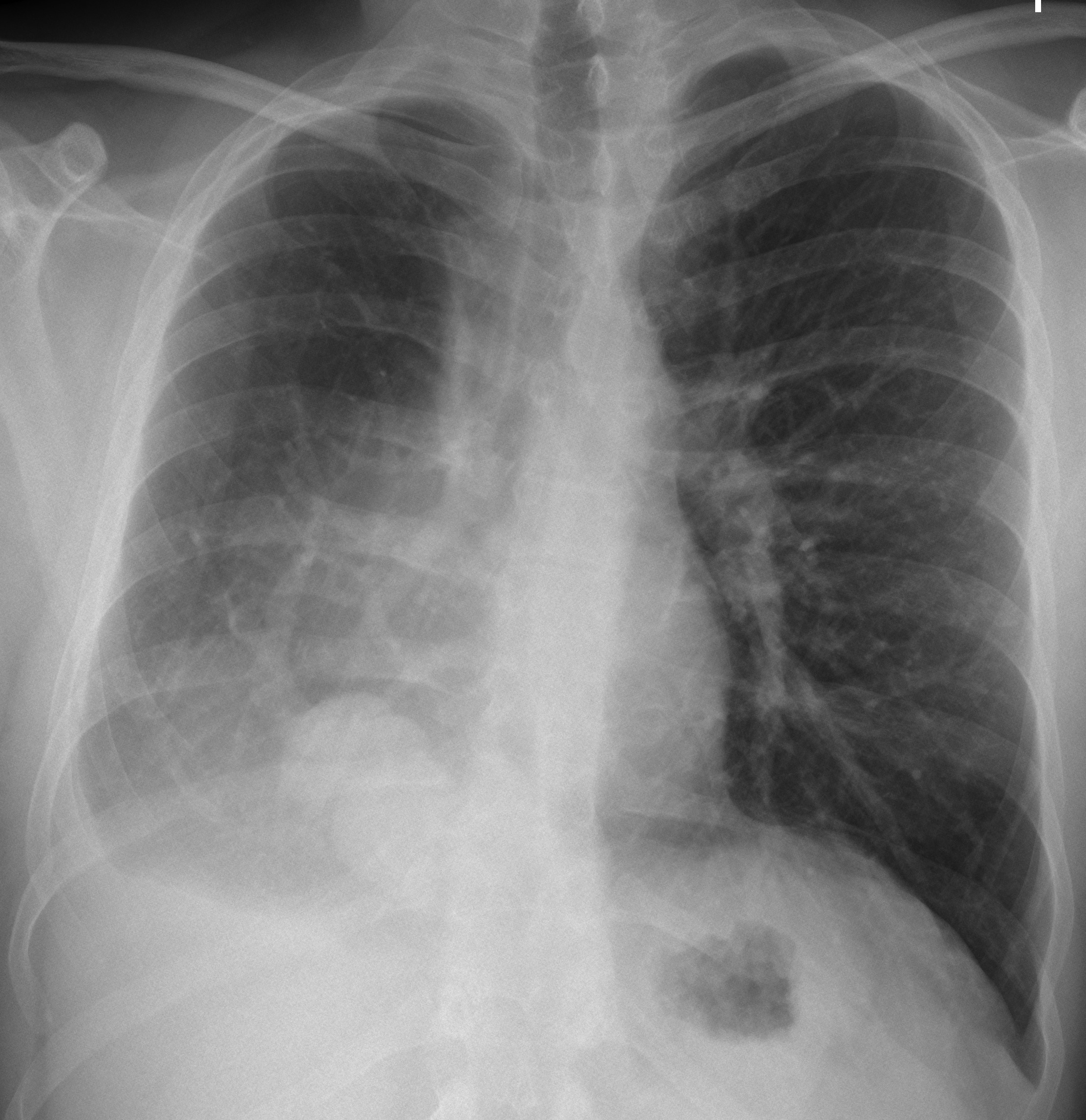
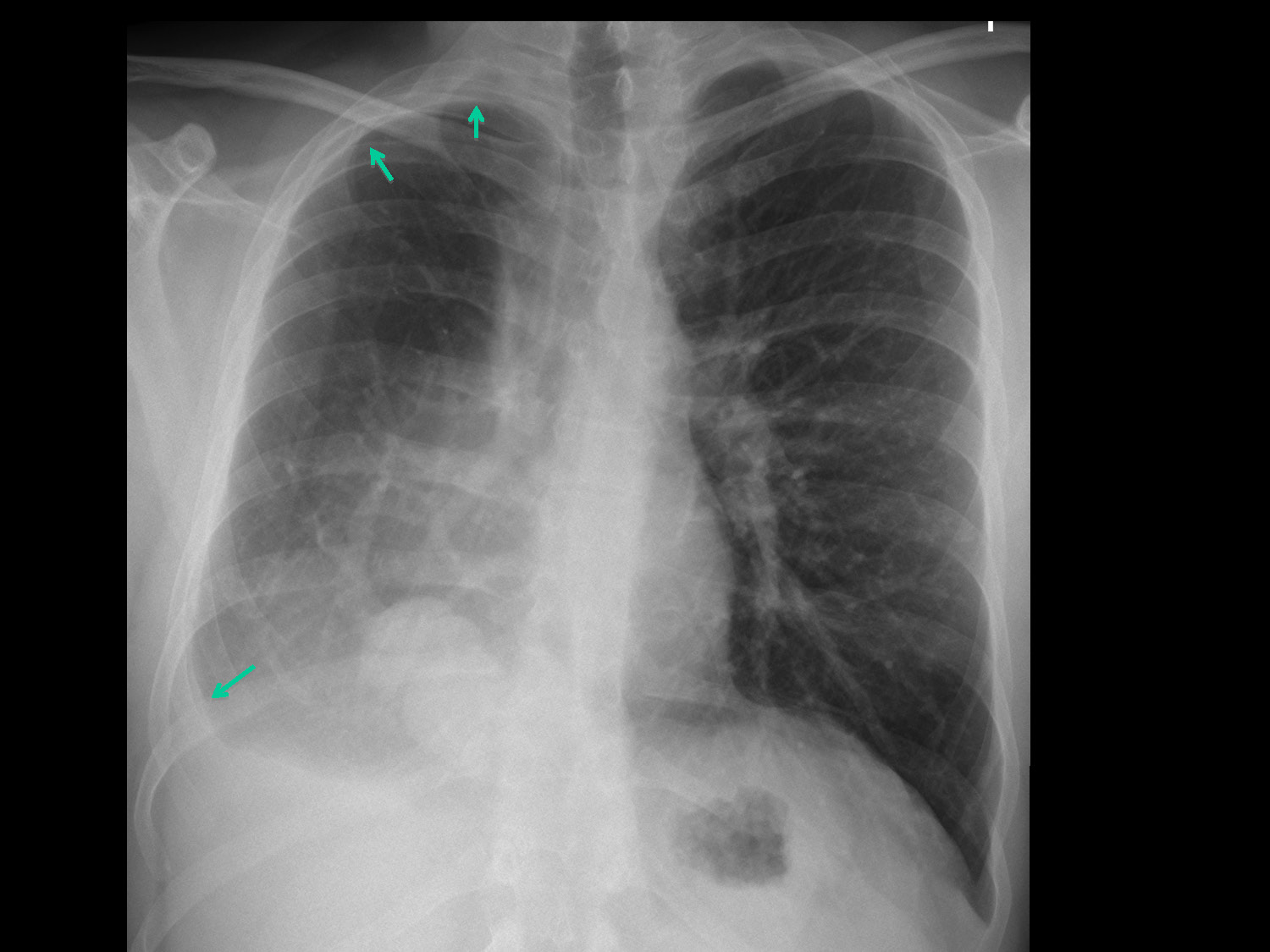
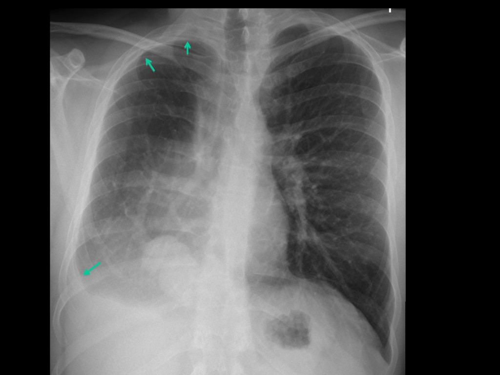
In the PA chest radiograph the right hemithorax is smaller than the left and there is apparent pleural thickening at the apex y right costophrenic angle (
arrows). There is slight displacement of the heart towards the right and blurring of the right heart border.
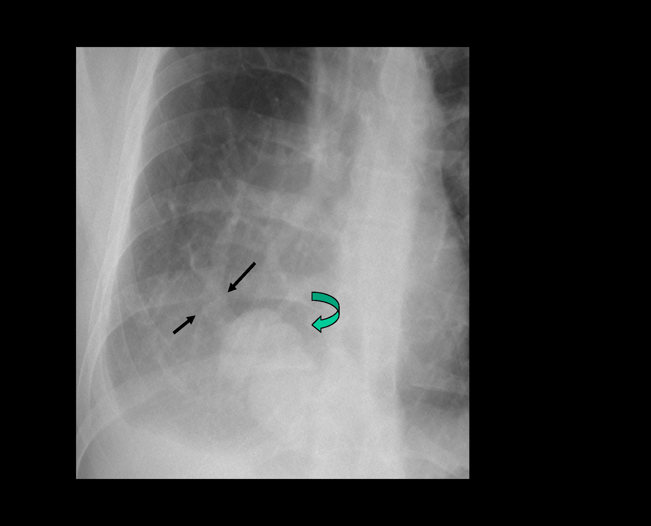
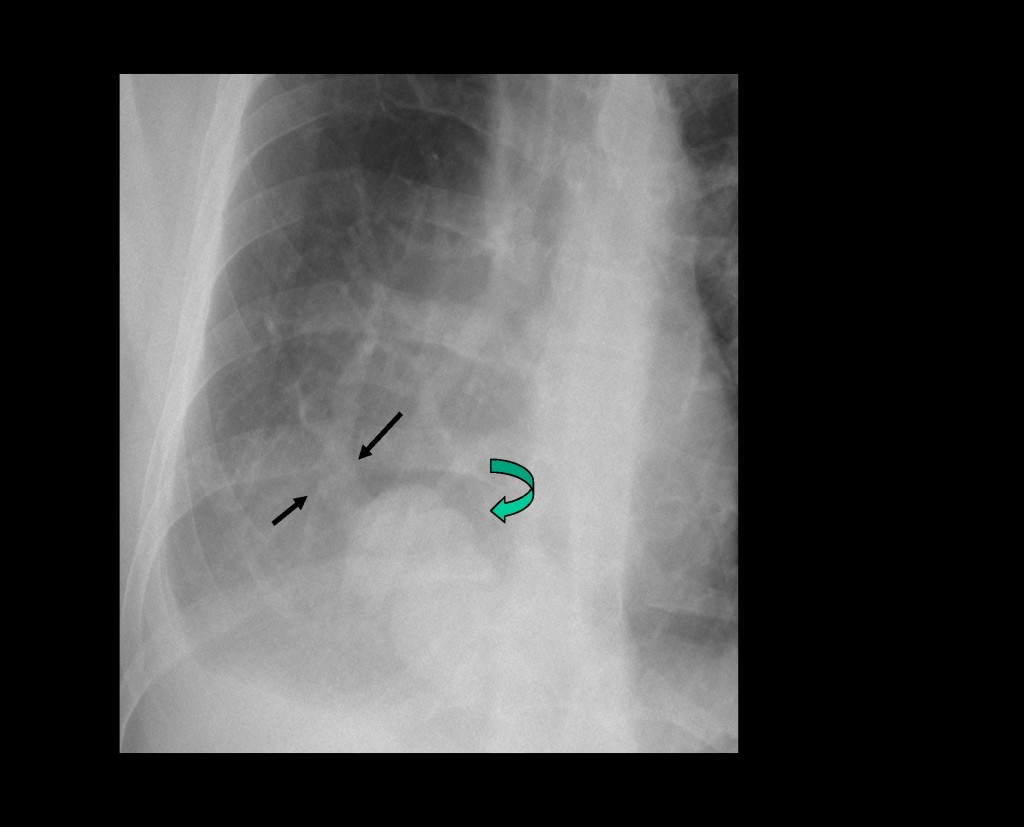
The cone down view shows a vessel cursing obliquely towards the inferior vena cava (black arrows) and a rounded shadow in the lower lung (curved arrow).
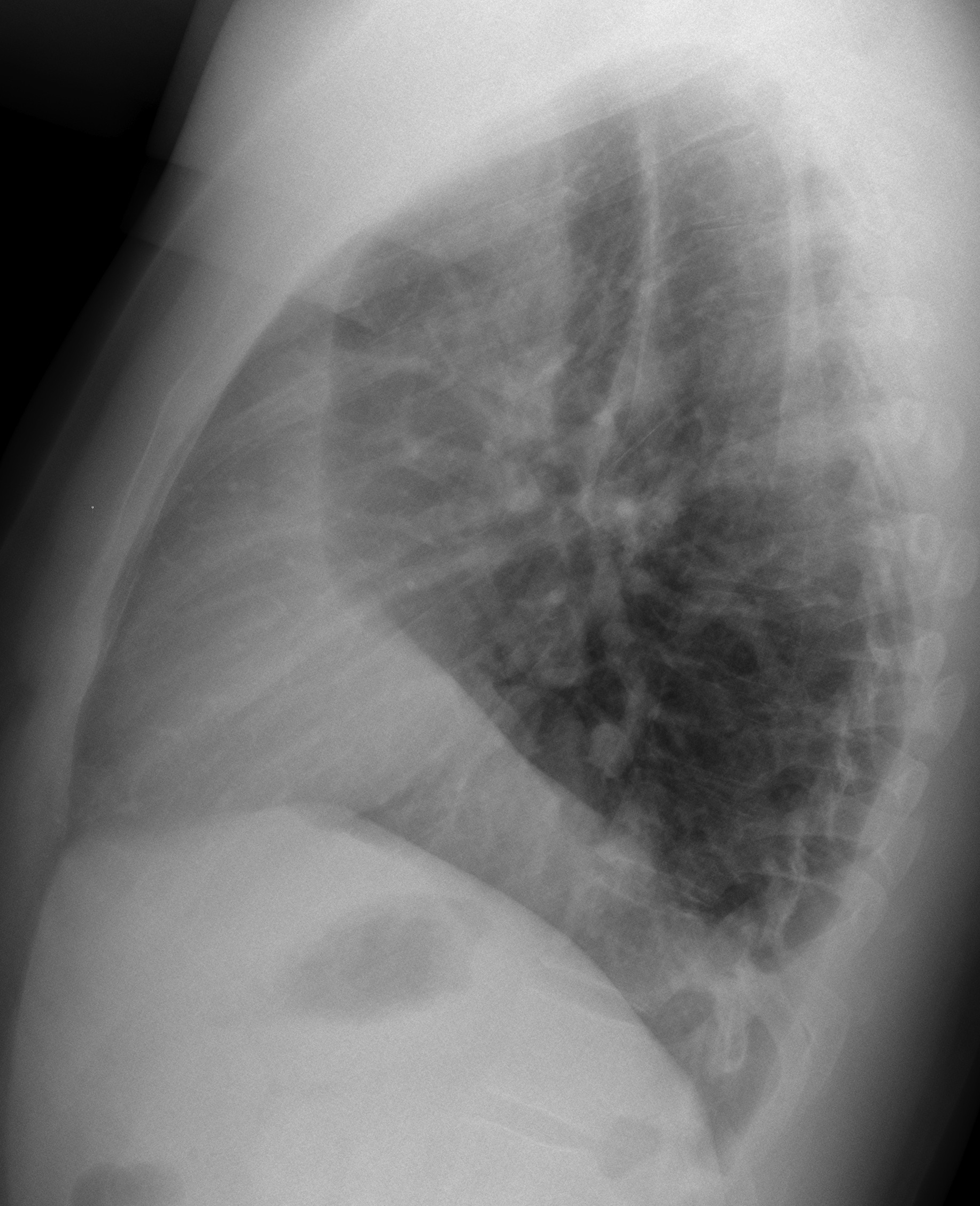
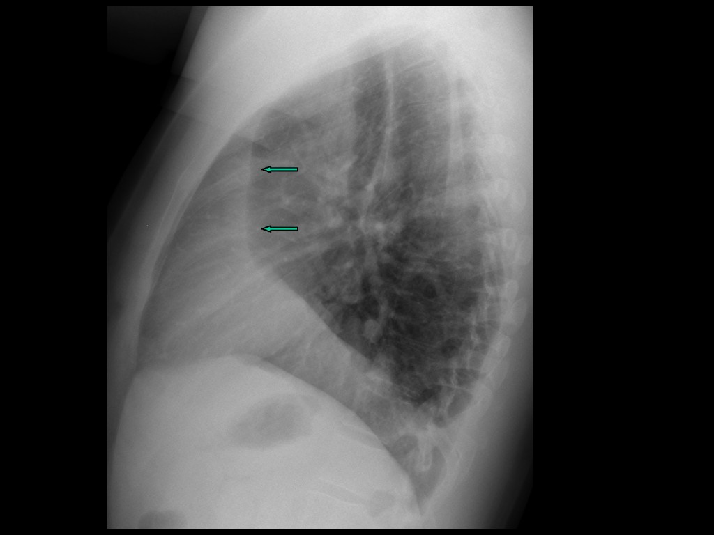
The lateral view shows a vertical retro-sternal band (arrows) and elevation of the right hemidiaphragm.
These findings are very suggestive of hypogenetic lung syndrome, secondary to congenital absence of one or two lobes of the lung. It almost always occurs on the right side. About 80% of the patients have anomalous venous drainage of the lower lobe (scimitar syndrome), which is visible in about half of them.
Sagital and coronal CT confirms the anomalous vein (A,B, green arrows). The apparent mass in the lower lobe (red arrows) is due to partial herniation of the liver (diaphragmatic defects are not unusual in this entity). Axial CT shows the small right lung and the abnormal bronchi (C, arrow). The apparent pleural thickening (D, arrow) is secondary to extrapleural fat, which increases and expands to compensate for the diminished lung volume.

Teaching point: congenital malformations are not unusual in adults. It is important to recognise them to avoid confusing them with other pathology.
Muppet adds: don’t rely only on seeing the anomalous vein. You have a 40/60 chance of seeing it!
 Dear friends,