Dear Friends,
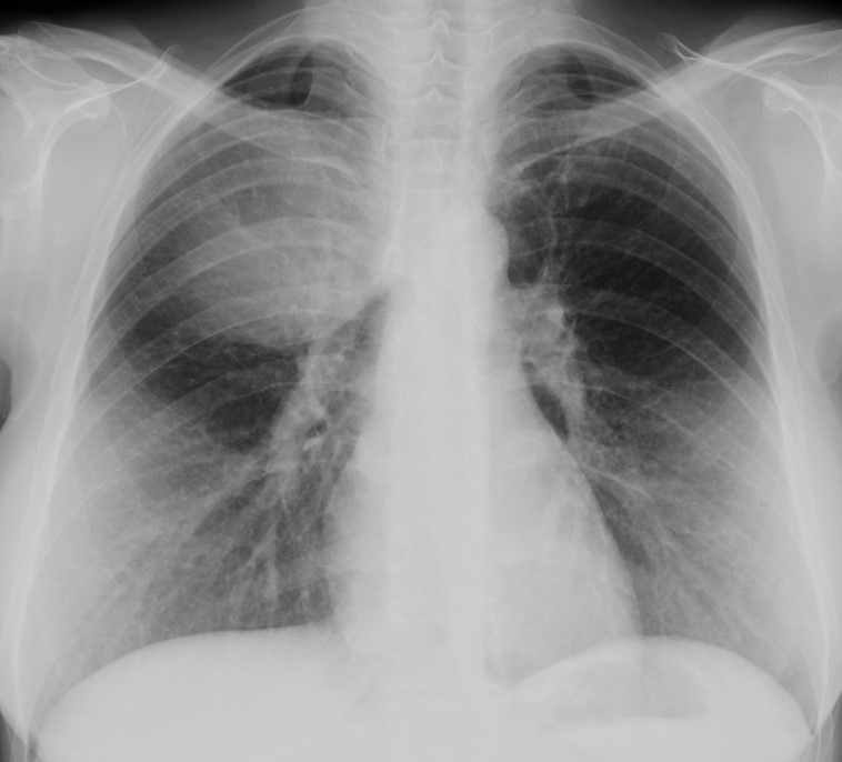
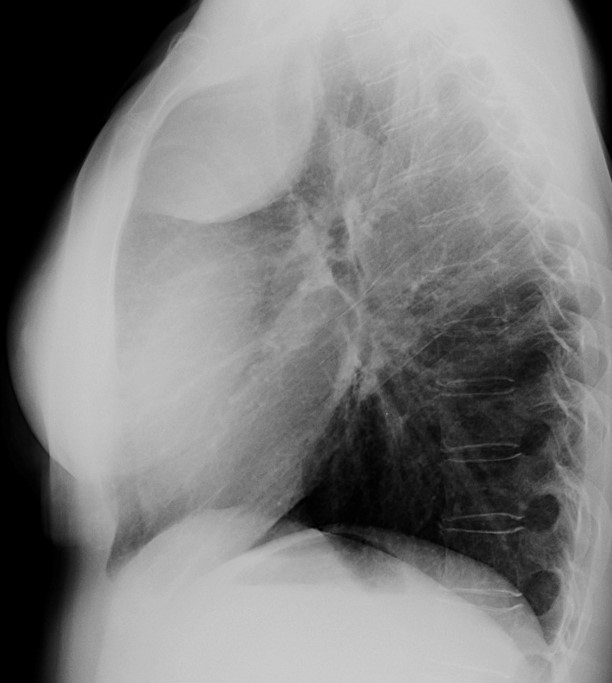
Muppet is feeling very guilty about the difficulty of case 100. To regain your sympathy, he has selected an easy case: radiographs belong to a 53-year-old woman with moderate pain in the right hemithorax for the last six months. Where is the lesion?
1. Lung
2. Mediastinum
3. Pleura/chest wall
4. Can’t tell
Check the images below, leave your thoughts in the comments sectiona and come back on Friday for the answer.
Click here for the answer to case #101
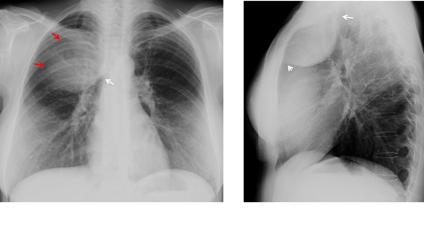
Findings: PA and lateral radiographs show an obvious rounded opacity projected over the right upper lung. In the PA view, the opacity has an acute angle with the mediastinum (arrow), which excludes a mediastinal origin. It is sharply defined medially and loses the border when contacting with the chest wall (incomplete border sign, red arrows). In the lateral view it has well-defined borders and obtuse angles at the extremes (arrows). The appearance is unequivocally extrapulmonary, placing the lesion in the pleura/chest wall compartment. No rib lesion is seen.
Coronal and sagittal CT show a soft tissue mass (arrows), without involvement of the ribs or chest wall. Patient was operated on and a pleural mass was found.
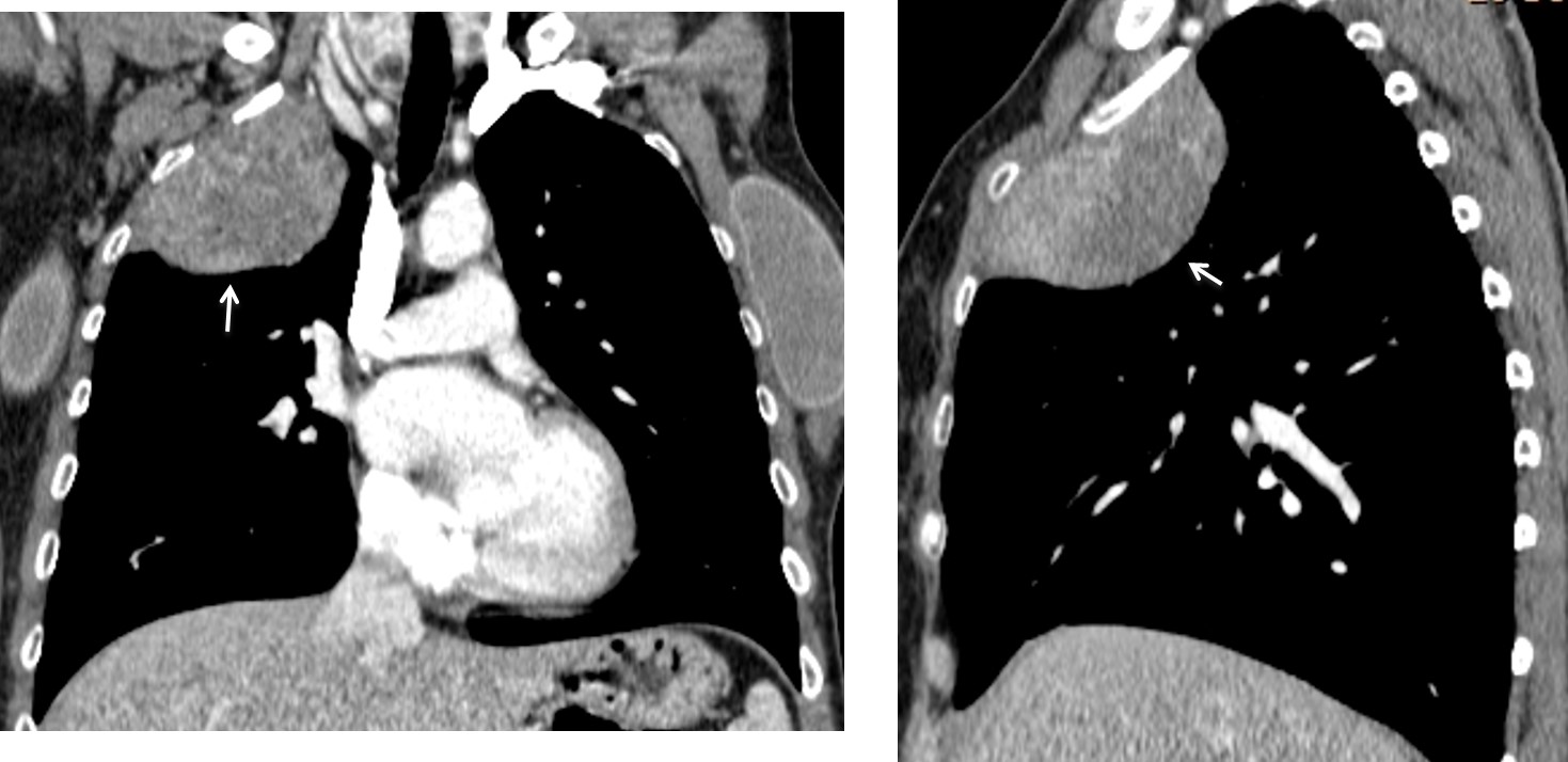
Final diagnosis: fibrous tumour of pleura
Most of you made the correct choice and demonstrated your knowledge of semiology. Must give credit to the first one, Dr. Elsayed Kotb
Teaching point: remember the basic signs of an extrapulmonary lesion: it has an obtuse angle at the ends and is sharply defined except when contacting with the chest wall.


