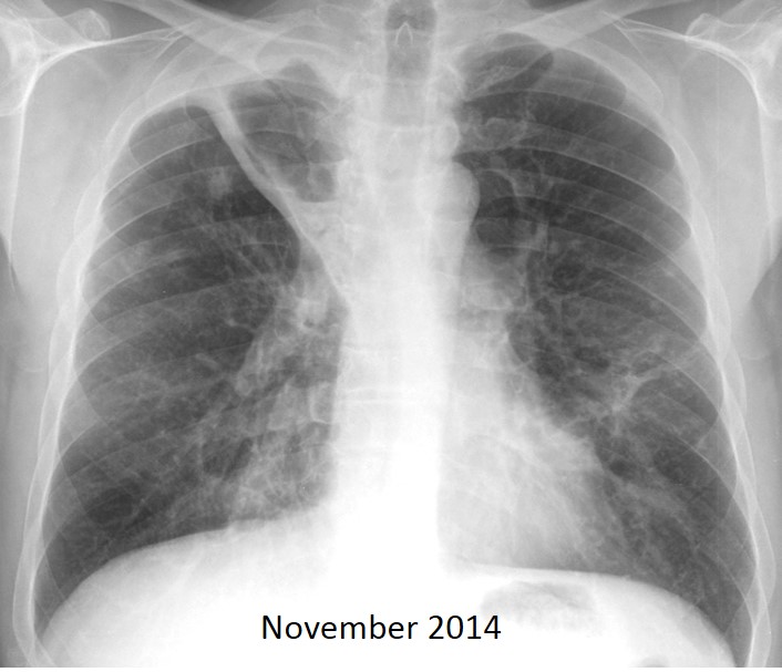
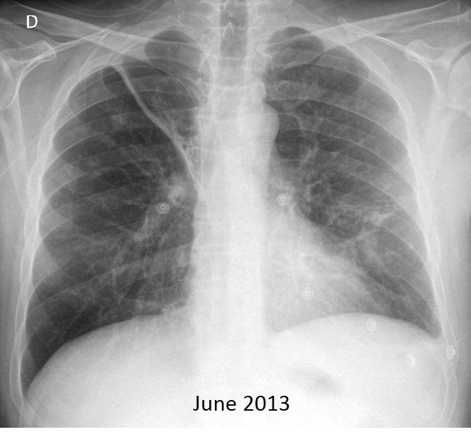
Today I am showing a recent case seen by my good friend Dr. Josep Mata. Radiographs belong to a 48-year-old man with haemoptysis and history of previous TB. Have a look at the images below, leave your thoughts and diagnosis in the comments section, and come back on Friday for the answer.
1. Infected bulla
2. Reactivation TB
3. Carcinoma in previous cavity
4. None of the above
Findings: PA radiograph in June 2013 shows a cavity in the RUL (A, arrow), with moderate peripheral pleural thickening (A, red arrow). One and a half years later, the wall of the cavity is thicker (B, arrow) and there is increased thickening of the pleura as well (B, red arrow).
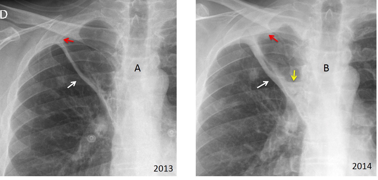
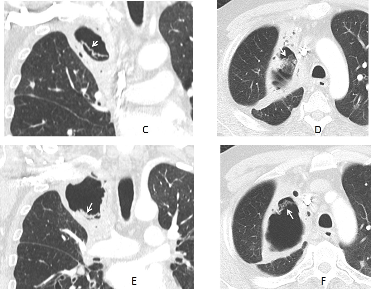
There is a small air-fluid level (B, yellow arrow). This progression is suspicious of aspergillus colonising a pre-existing cavity. CT demonstrates spongiform tissue of fungal strands inside the cavity (C-F, arrows).
Final diagnosis: Mycetoma
Congratulations to Nicky, who was the first to suggest mycetoma.
Teaching point: remember that progressive pleural thickening in the periphery of a pre-existing cavity is an early indicator for mycetoma.