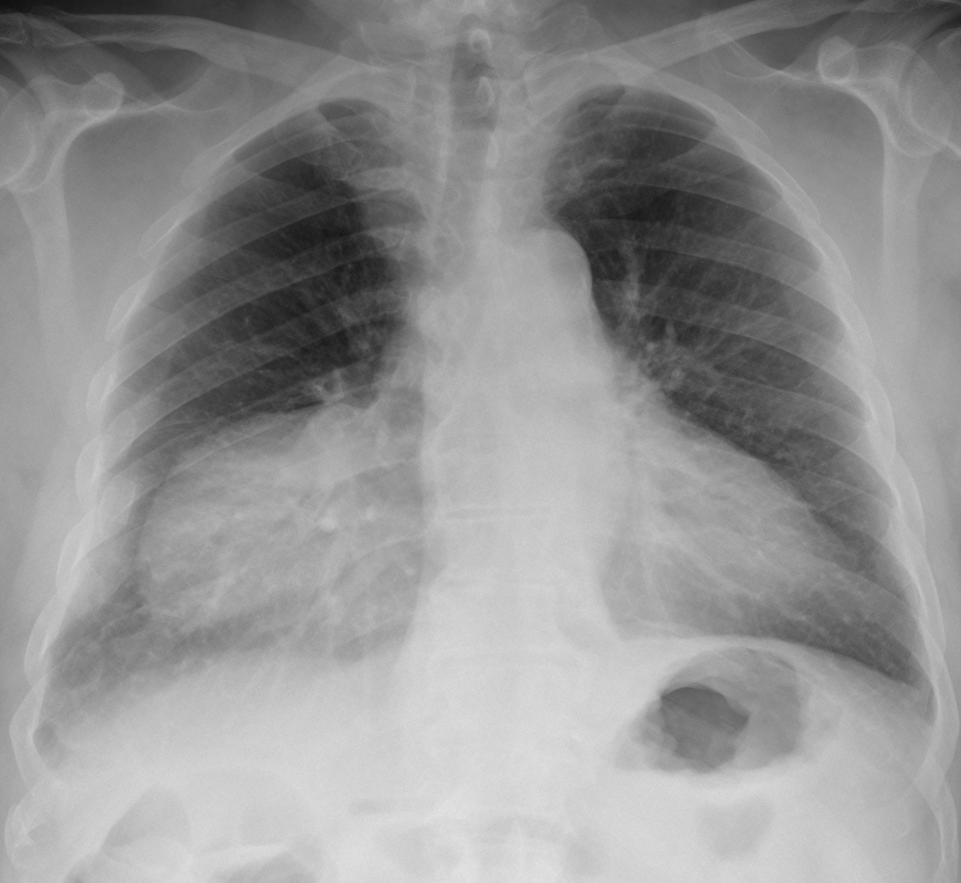
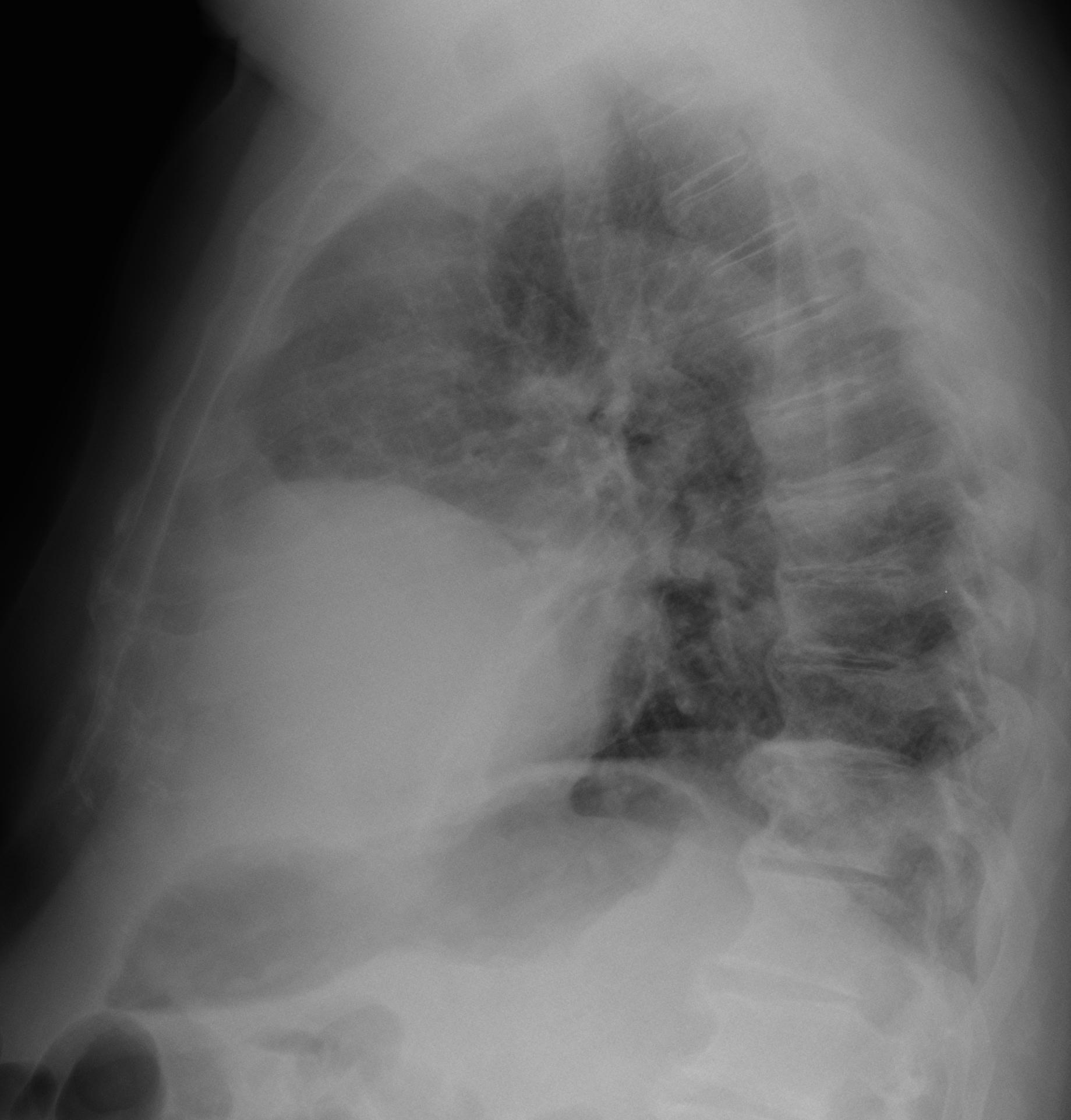
Muppet is in Naples this week and left me alone to present the case of a 58-year-old man, asymptomatic. Check the images below, leave me your thoughts and diagnosis in the comments section, and come back on Friday for the answer.
1. Epicardial fat pad
2. Morgagni’s hernia
3. Pericardial cyst
4. None of the above
Findings: PA and chest radiographs show a well-defined mediastinal mass at the right cardiophrenic angle (A,B arrows).
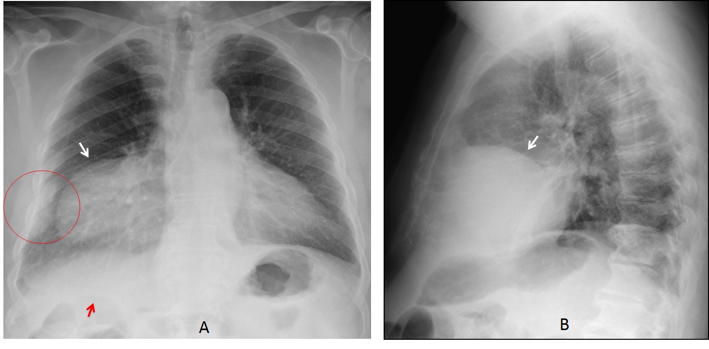
The appearance of the mass is unspecific but there are two clues to a possible diagnosis: there is blunting of the right costophrenic angle accompanied by two consolidated costal fractures (A, circle) and the splenic angle of the colon (A, red arrow) is too high, indicating a small liver. Believe it or not, these two findings raised the possibility of a traumatic diaphragmatic hernia, with part of the liver above the diaphragm.
Review of the patient´s history revealed a car accident in 1988 with diaphragmatic rupture. A follow-up CT clearly shows the diaphragmatic rupture (C,D, arrows) and the herniated liver.
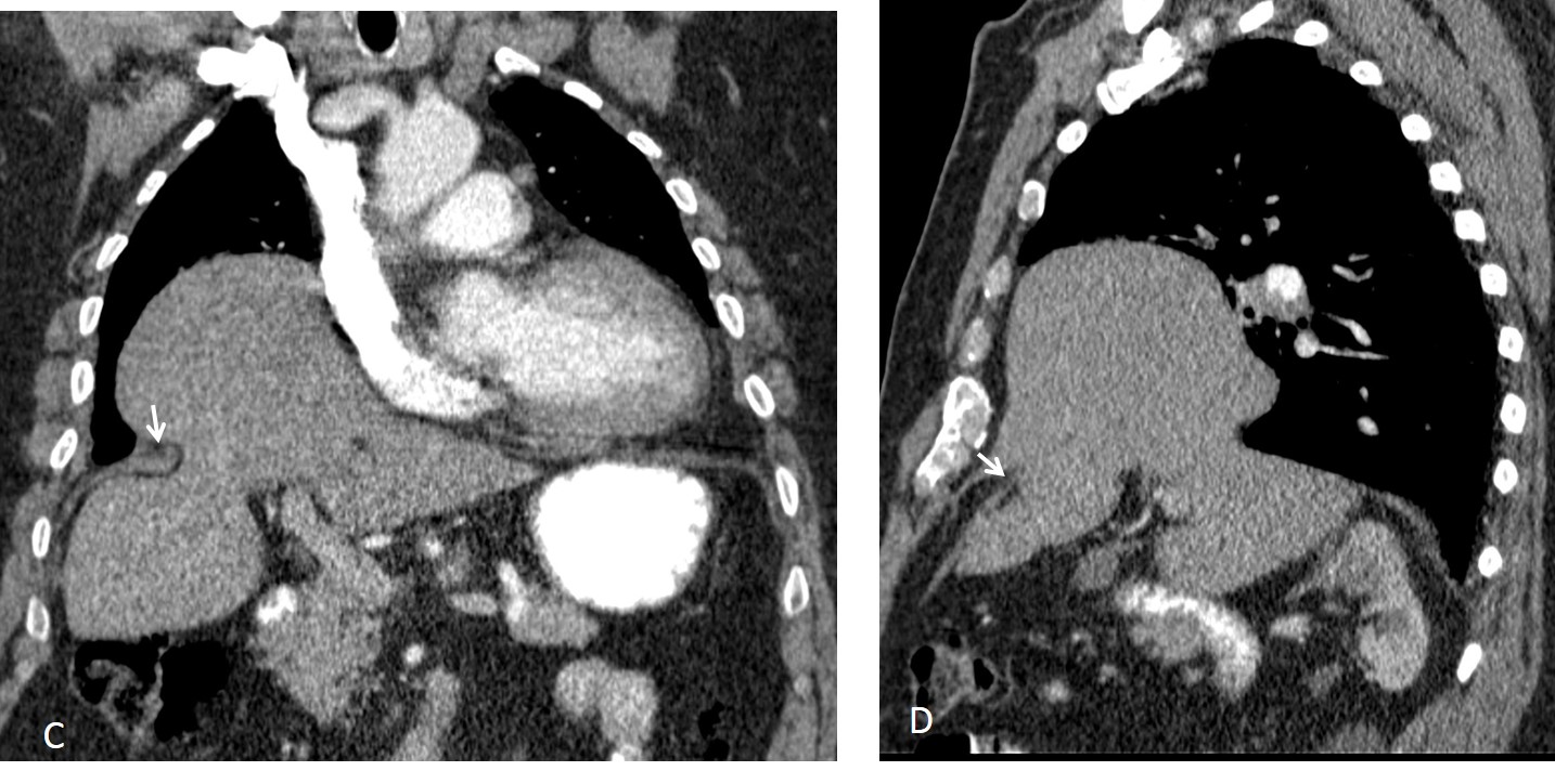
Final diagnosis: partially herniated liver, secondary to traumatic diaphragmatic hernia.
Congratulations to Genchi Bari and Nikos, who made the correct diagnosis
Teaching point: Remember that any chest opacity adjacent to the diaphragm may arise from the abdomen. It worked in this case!