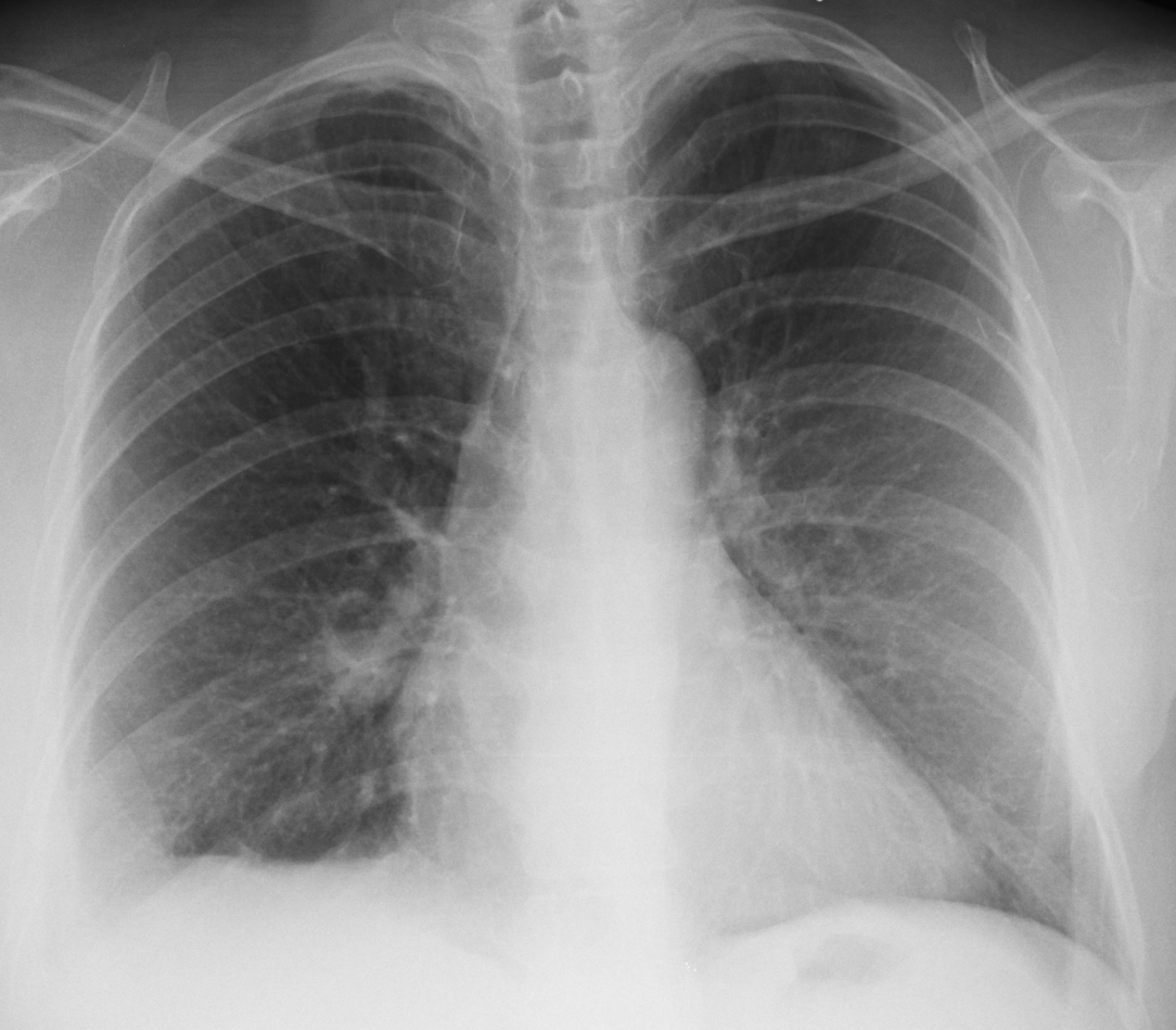
Dr. Pepe has eloped to the Bahamas with Miss Piggy and forgot to prepare the Diploma case. Hope he returns happy and suntanned. In the meantime I will show images of a 57-year-old woman with acute chest pain and mild fever. Check the images below, leave your thoughts and diagnosis in the comments section and come back on Friday for the answer.
1. Pneumonia
2. Pulmonary infarct
3. Pleural fluid
4. None of the above
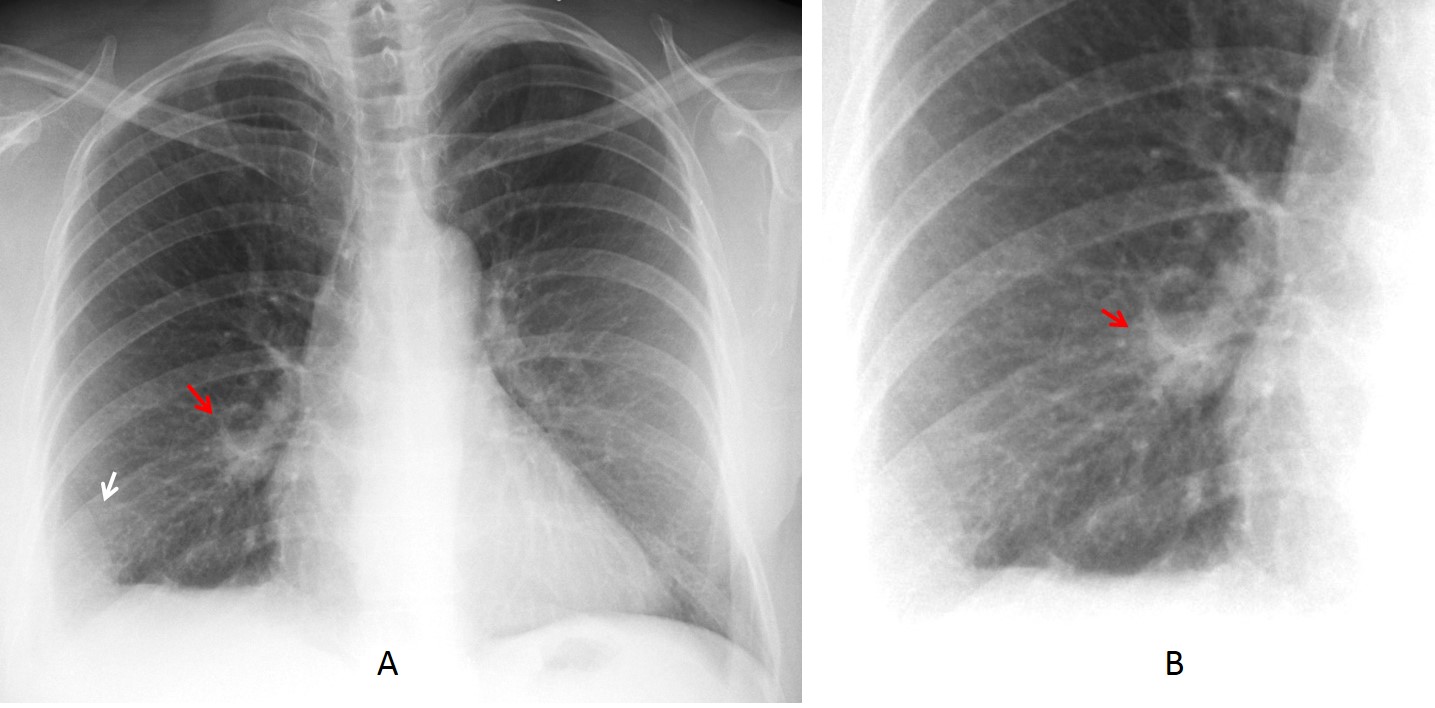
Findings: PA radiograph shows a triangular pulmonary opacity at the right costophrenic angle (A, arrow), with the characteristic appearance of a Hampton hump, suggesting a pulmonary infarct. In addition, a round nodule is visible in the lower aspect of the right hilum (A,B red arrows).
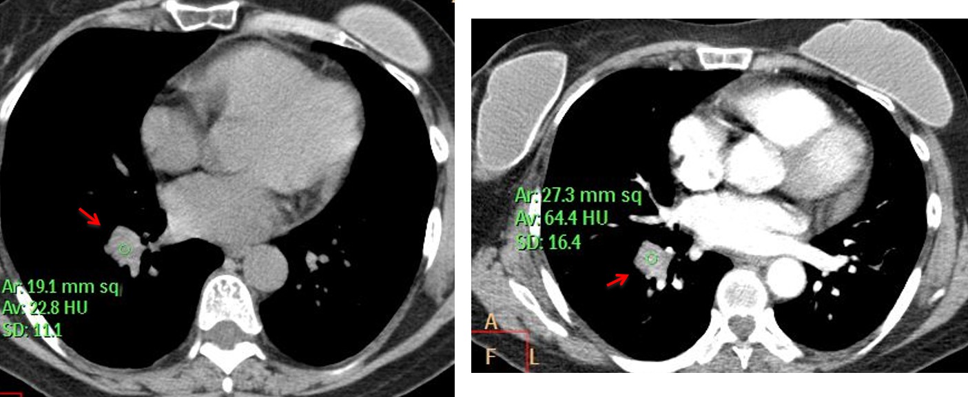
CT confirmed the pulmonary embolism and a RLL mass (C,D arrows) that enhanced 42 H.U. after contrast injection, suggesting malignancy. Needle biopsy confirmed a lung adenocarcinoma.
Final diagnosis: Carcinoma of the lung with pulmonary infarct.
Congratulations to Dr. Prasanna Kulkarni who made the correct diagnosis
Teaching point: remember avoiding satisfaction of search. In this case, it discovered an unsuspected malignancy, which is known to increase the risk of pulmonary embolism.