
Dear Friends,
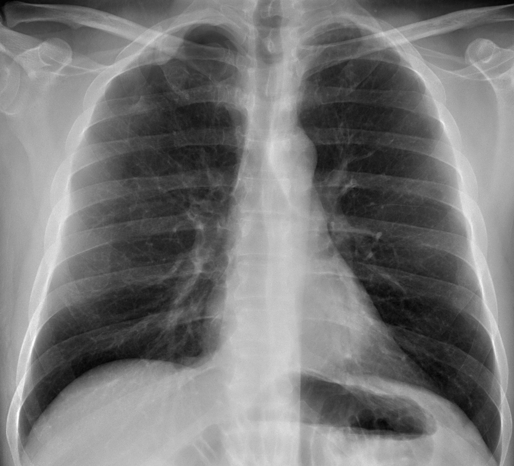
After the ECR, we all deserve an easy case. Showing routine control radiographs of a 51-year-old man operated on for synovial sarcoma of his right leg fourteen years ago.
Do you see any abnormality?
Check the images below, leave your thoughts in the comments section and come back on Friday for the answer.
Click here for the answer to case #111
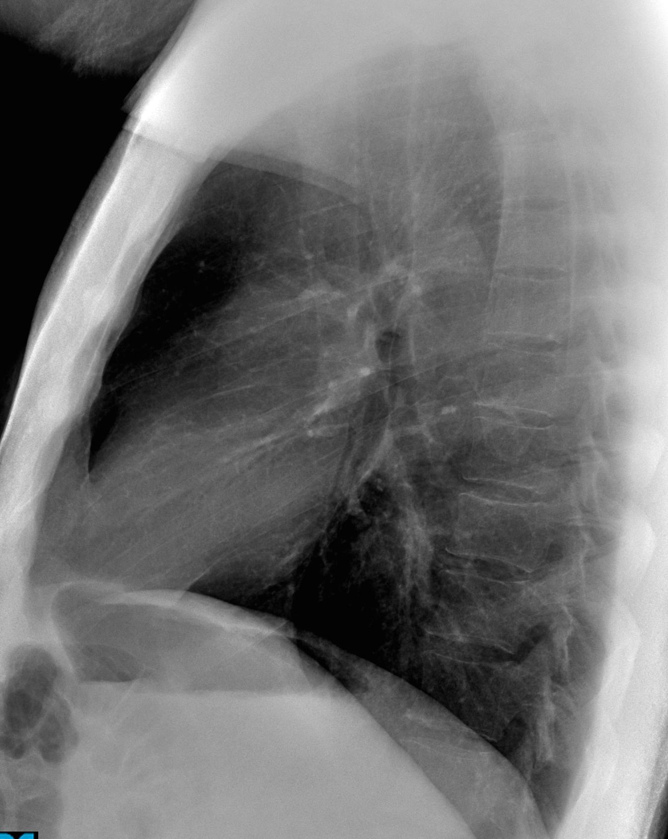
Findings: PA radiograph shows an opacity in the right apex with the typical appearance of an extrapulmonary lesion (A, arrow). It is seen better in the cone down view, which also shows a pneumothorax (B, arrows), secondary to percutaneous biopsy. The lateral view is unremarkable.
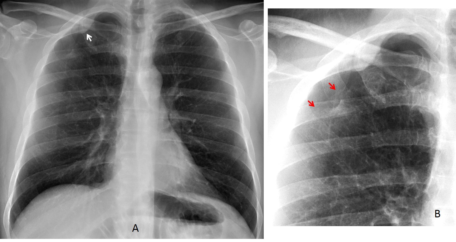
CT and MRI confirm a parietal mass (C,D, arrows). At surgery, a neurogenic tumour of the chest wall was found.

Final diagnosis: malignant schwannoma
Congratulations to j.d. who discovered the mass.
Teaching point: I am showing this case because it is an excellent example of extrapulmonary lesion: well defined and with tapering margins.


