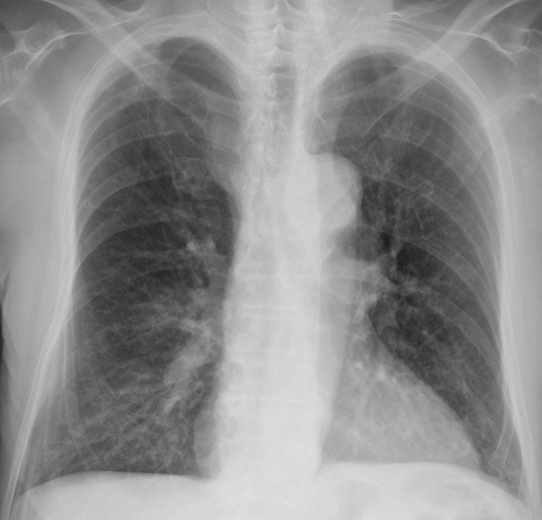
Muppet came back in a good mood and wishes to present an easy case. Radiographs belong to a 73-year-old male smoker with a moderate cough. What do you see?
Check the images below, leave your thoughts in the comments section and come back on Friday for the answer.
Findings: PA chest shows a mediastinal mass protruding at the level of the aorto-pulmonary window (Fig 1A, arrow). The mass is also evident in the lateral view (Fig 1B, arrow).
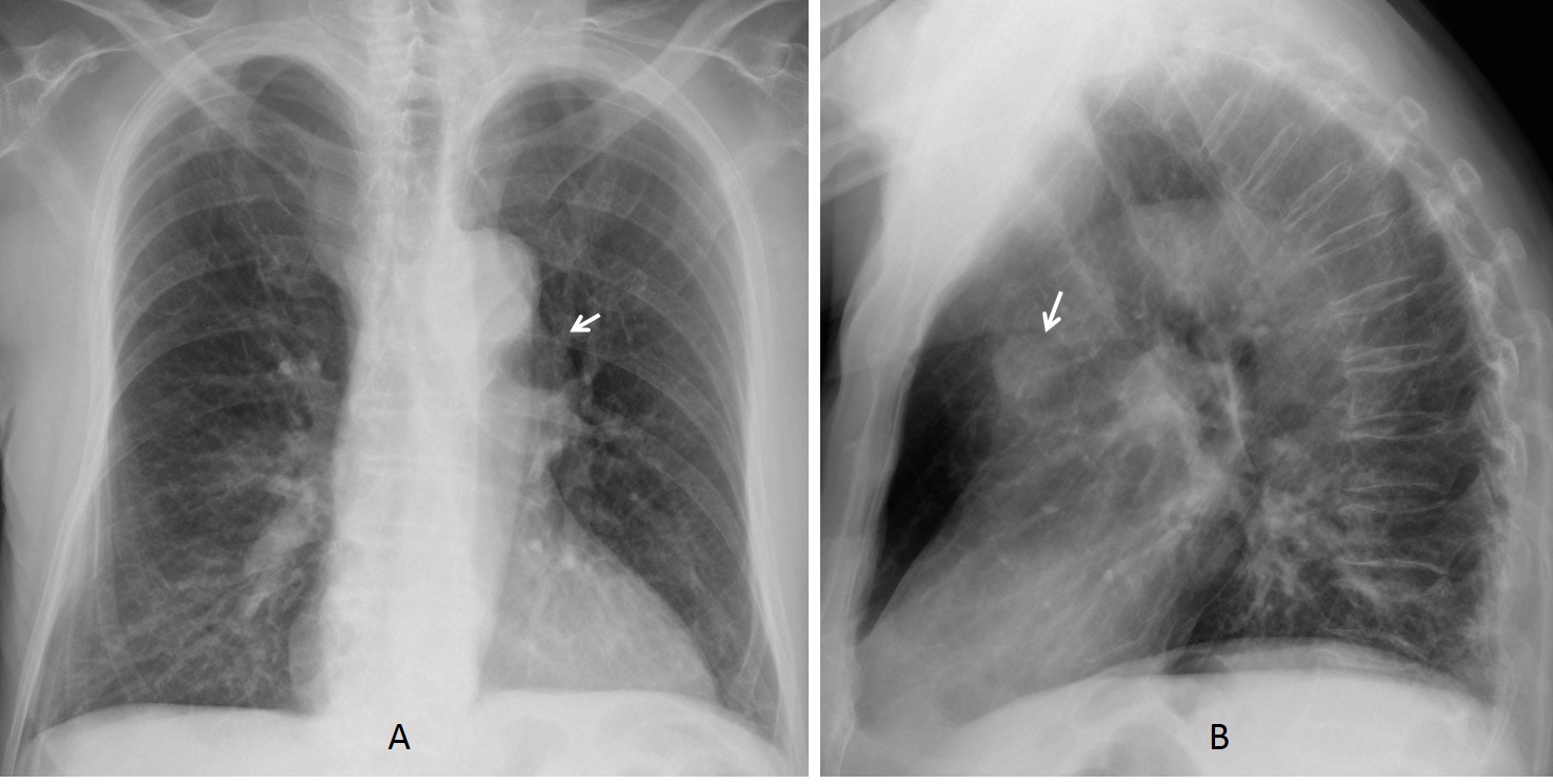
Fig. 1
Radiographs taken five years earlier did not show the abnormality (Fig 2).
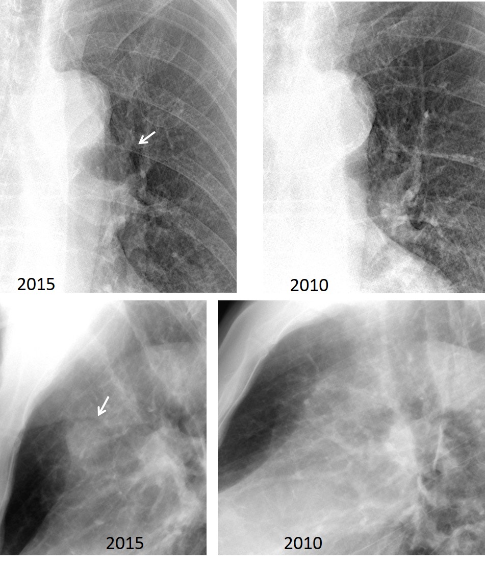
Fig. 2
The location of the lesion suggests enlarged lymph nodes as the first possibility, although other etiologies cannot be excluded. Enhanced axial and coronal CT demonstrates that the mass represents a saccular aneurysm arising from the aortic arch (Fig 3, arrows).
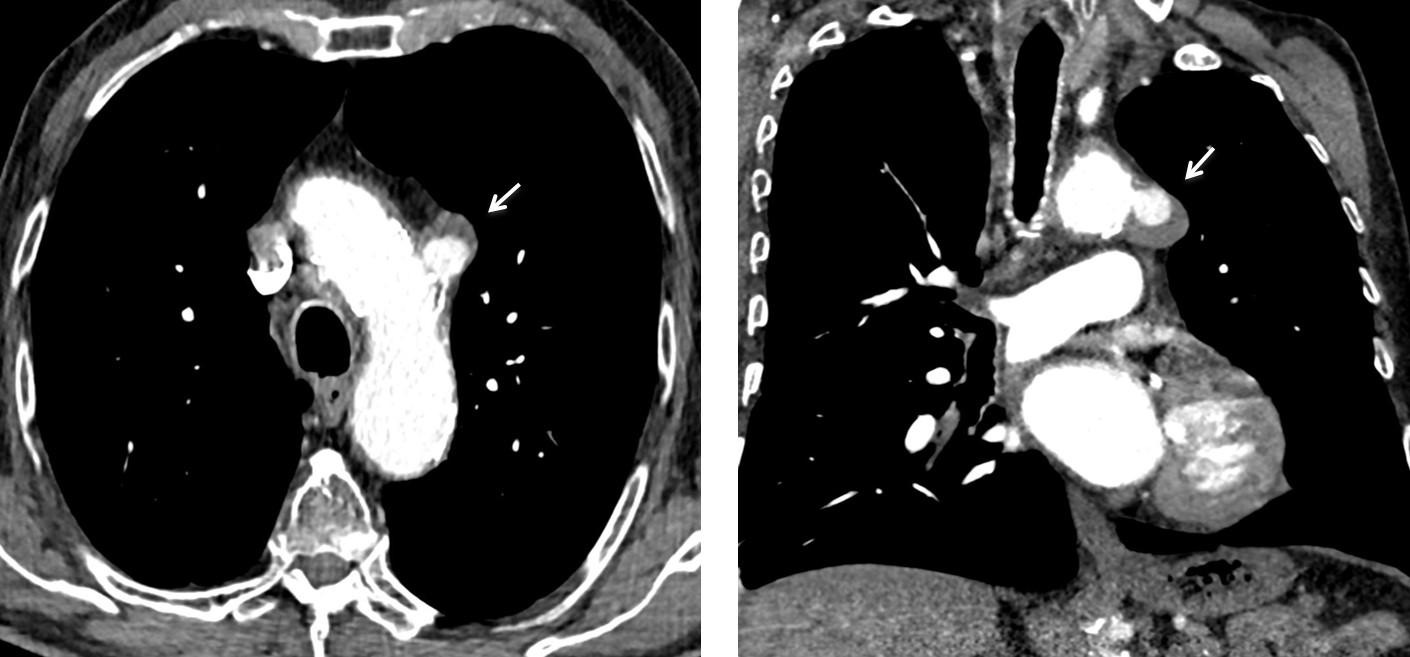
Fig. 3
Final diagnosis: saccular aneurysm of aortic arch.
Congratulations to Gus, who was the first to discover the abnormality
Teaching point: remember that any mediastinal mass may have a vascular origin. Enhanced CT should always be performed to explore this possibility.