Dear Friends,
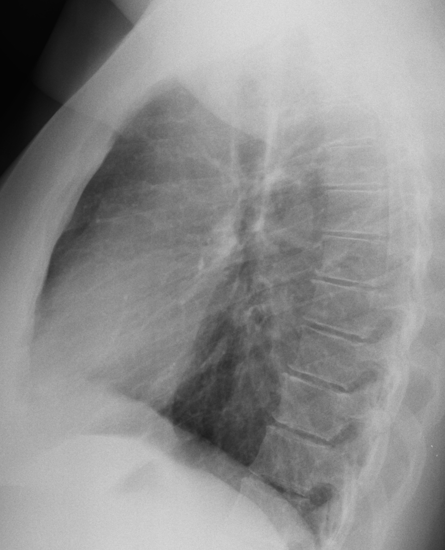
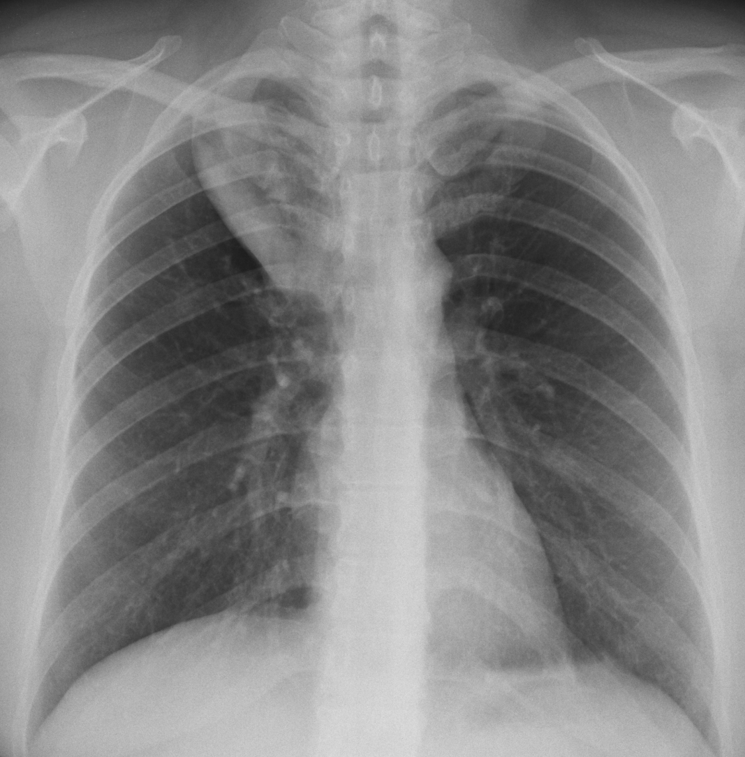
My good friend and former resident Romá Vidal sent me some images of a 39-year-old man with cough and moderate fever. Have a look at the images below, leave your thoughts in the comments section, and come back on Friday for the answer.
Diagnosis:
1. TB
2. RUL collapse
3. Fungus ball
4. None of the above
Click here for the answer to case #115
Findings: the lateral view is unremarkable. PA radiograph shows a well-defined triangular opacity in the right upper lung that at first look simulates an RUL collapse with a bulging fissure. The fact that the trachea is in the midline and the right hilum is not elevated go against this possibility. The clue to the diagnosis lies in recognising an upper trigonum (A, arrow) and a fissure (A, red arrow), which are the hallmark of an azygos lobe. The findings can be explained by an inflammatory process within the azygos lobe, simulating an RUL collapse.
A follow-up radiograph a month later shows the disappearance of the pneumonia and confirms the azygos lobe (B, arrow).
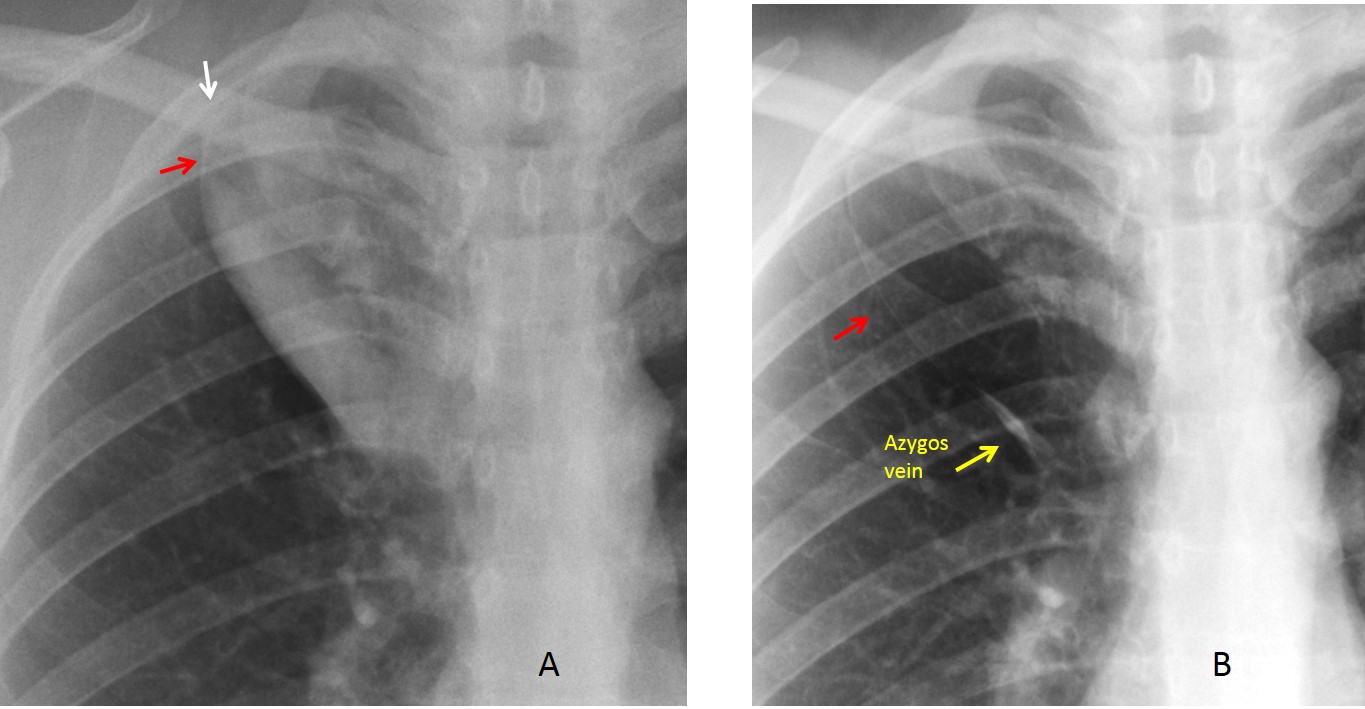
Final diagnosis: azygos lobe pneumonia
Congratulations to Fares, who was the first to mention the right diagnosis, with Lola la Piconera and Cocotet as close seconds.
Teaching point: remember that pneumonias have ill-defined margins except when limited by a fissure.


