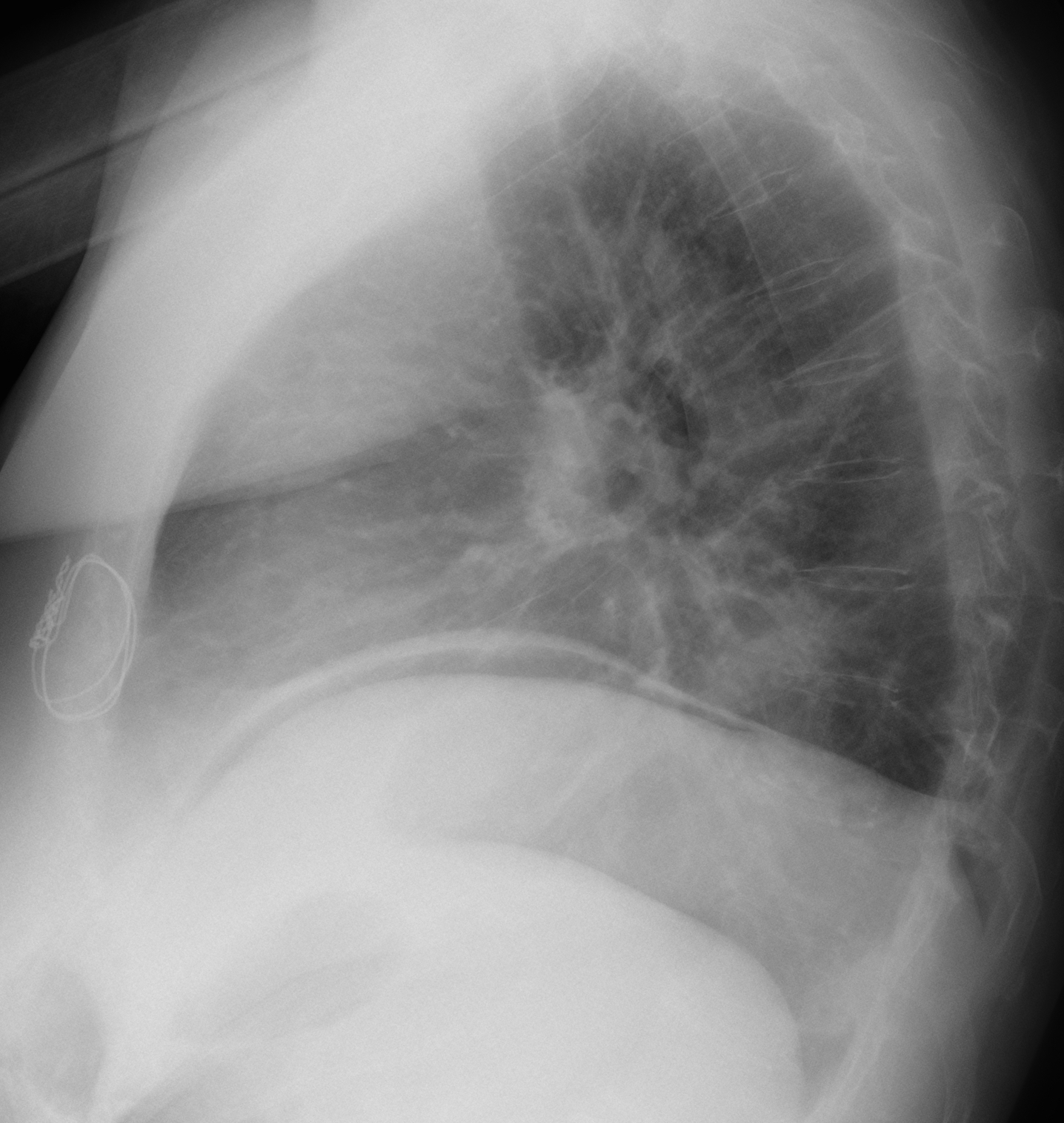
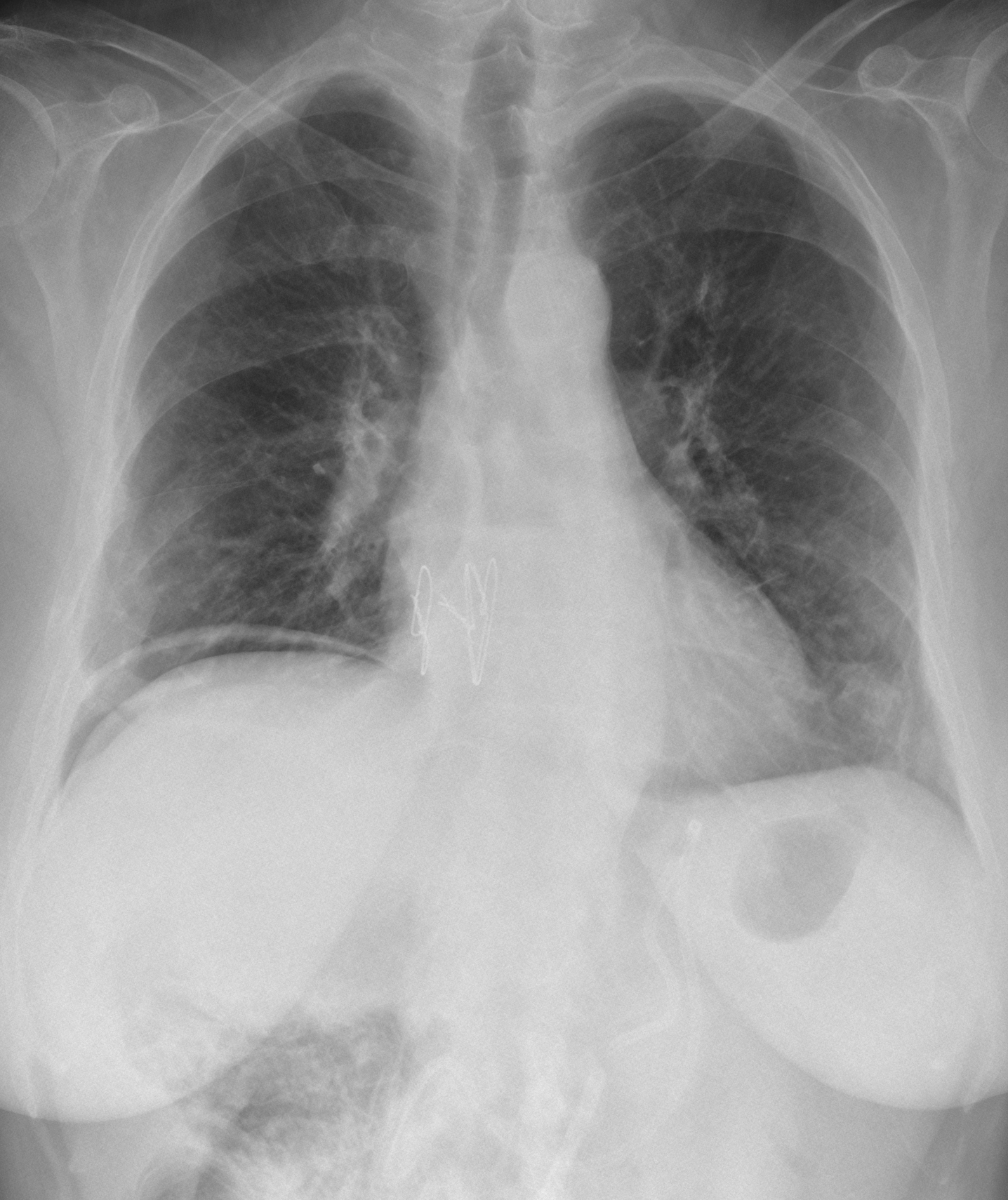
Since summer is coming (Game of Thrones revisited), I am presenting an easy case today. Images belong to an asymptomatic 67-year-old female with scleroderma. She had a lung transplant last year and a gastrostomy one month ago.
Check the images below, leave your thoughts in the comments section and come back on Friday for the answer.
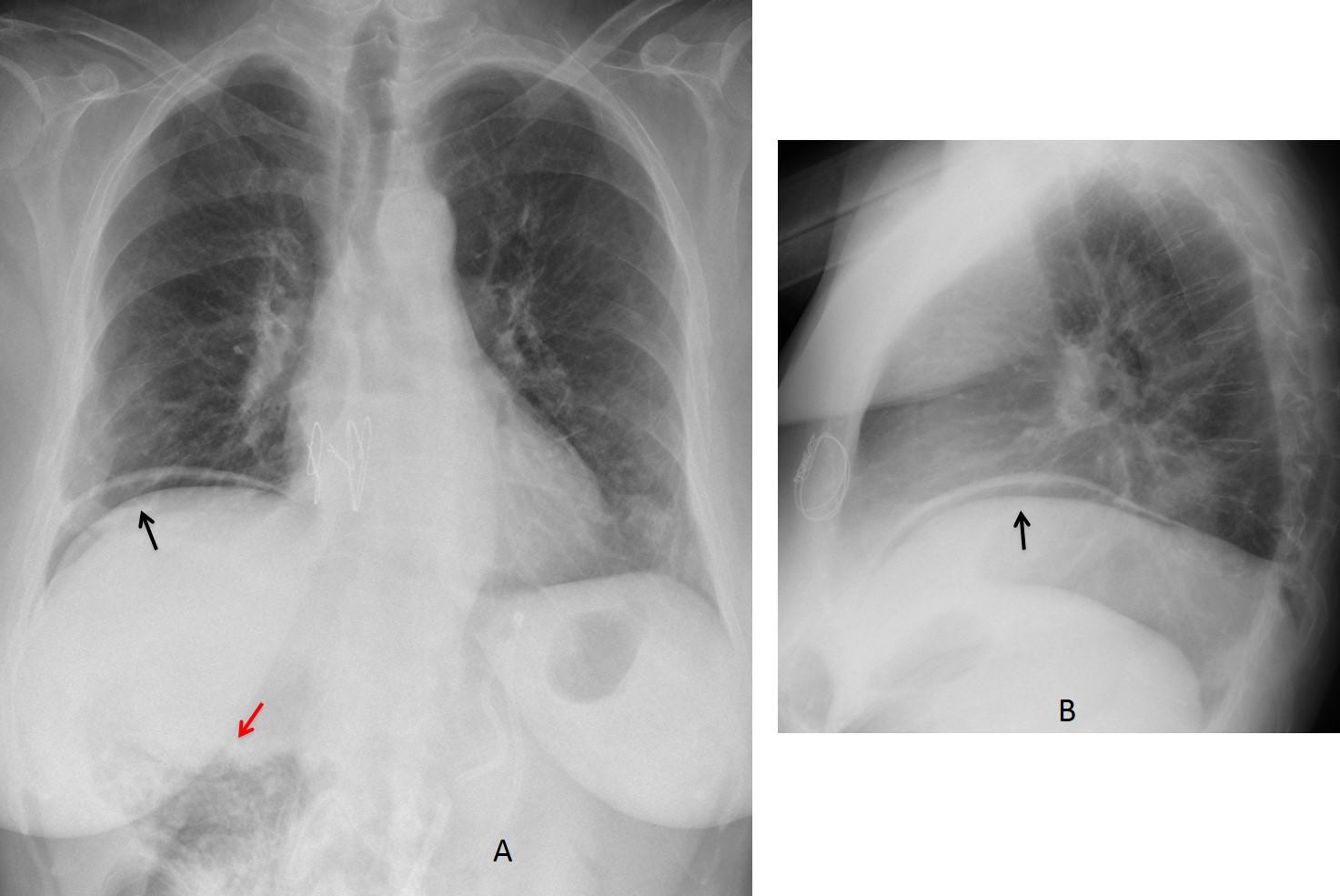
Findings: there is free air under the right hemidiaphragm (A-B, arrows). There is also a bubbly appearance of the hepatic flexure of the colon (A, red arrow), which suggests intestinal pneumatosis.
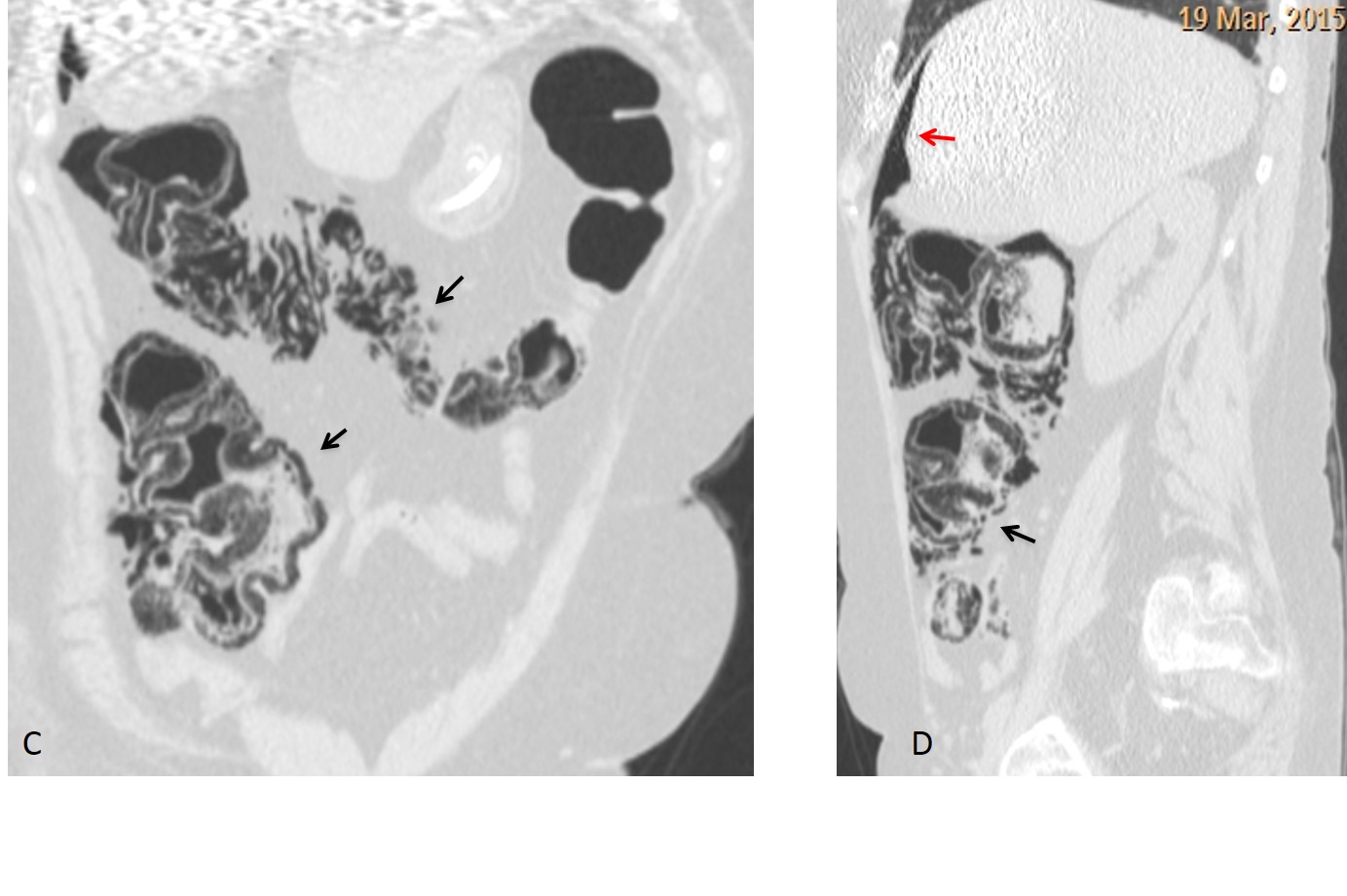
CT confirms pneumatosis of the right colon (C-D, arrows) and the free air (D, red arrow).
As the patient’s symptoms were minimal and scleroderma is known to cause pneumatosis, it was decided to wait. Four days later the pneumoperitoneum disappeared and the appearance of the hepatic flexure had improved (F, arrow).
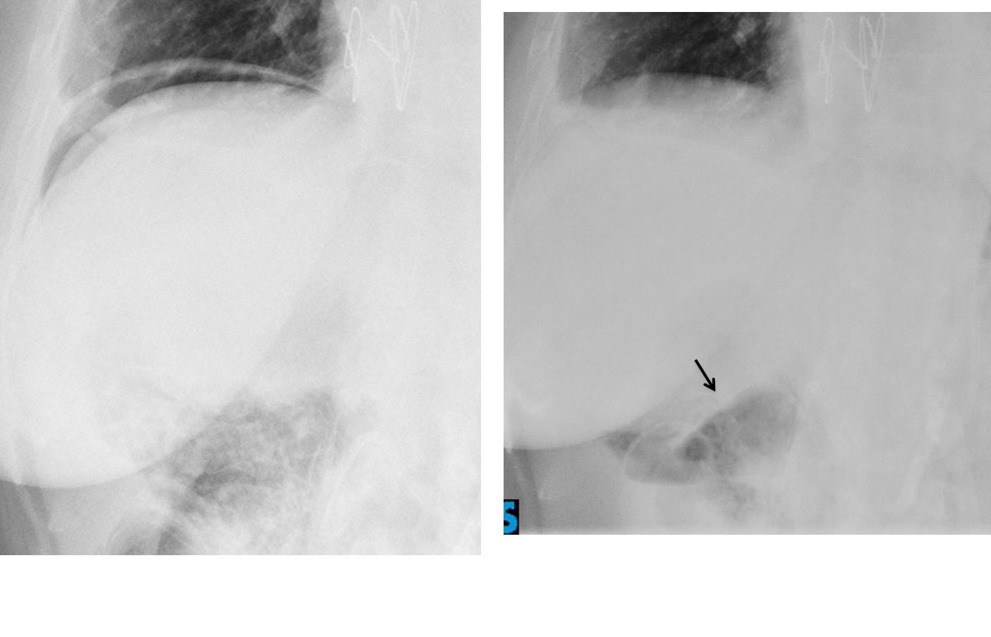
Final diagnosis: pneumatosis cystoides intestinalis with pneumoperitoneum, probably secondary to scleroderma.
Congratulations to Fares, who mentioned the right diagnosis.
Teaching point: remember that free abdominal air is either due to instrumentation or perforation of a hollow viscus. Intestinal pneumatosis should always be considered, especially if the appearance of the bowel suggests it.