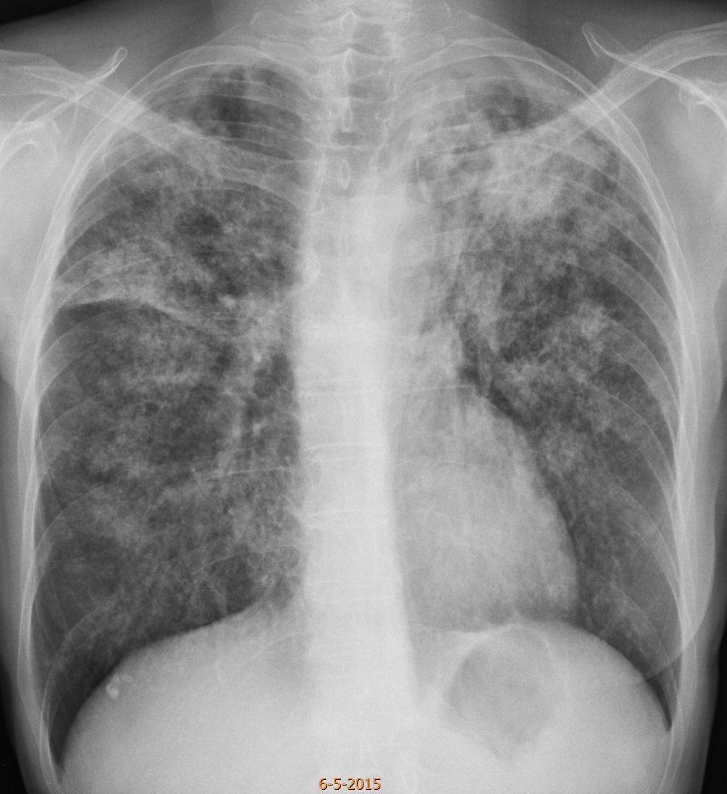
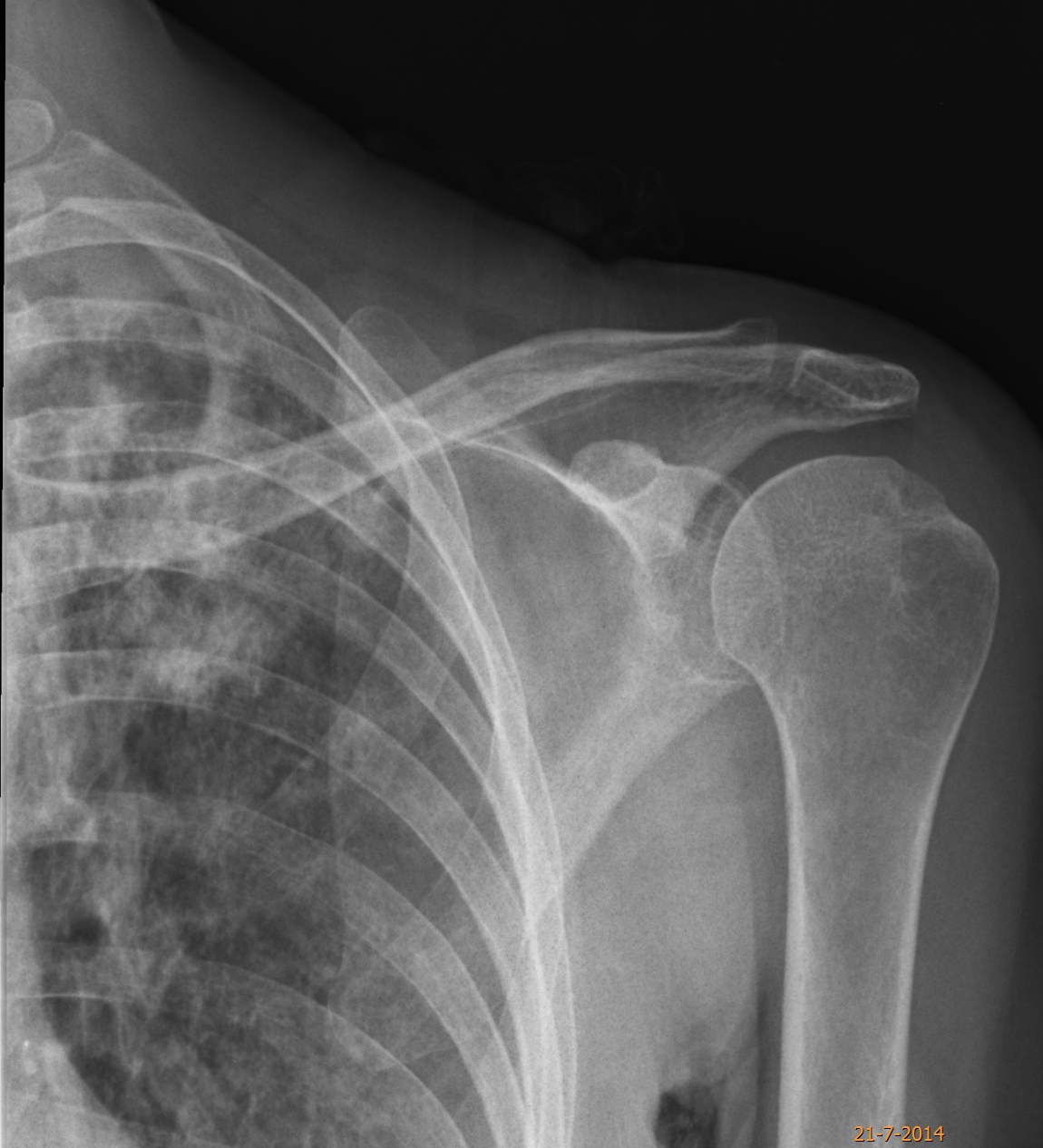
Welcome back to your favourite corner! Starting with a warm-up case: pre-op radiograph prior to biopsy of a vocal cord nodule in a 57-year-old woman with hoarseness. Including a radiograph of the shoulder taken eight months earlier in which part of the left lung is seen.
Check the images below, leave your thoughts in the comments section, and come back on Friday for the answer.
1. Widespread adenocarcinoma
2. TB
3. Sarcoidosis
4. None of the above
Findings: PA chest radiograph shows widespread air space disease located mainly in the upper lungs. Comparing with the previous film, the disease has progressed moderately in the last months. In my opinion, the clue to the diagnosis lies in detecting cavitation, depicted as radiolucent areas in both apices (A-B, arrows).
The fact that the one in the left apex has not changed (A-B, red arrows), despite the progression of the disease, supports the presence of cavitation.
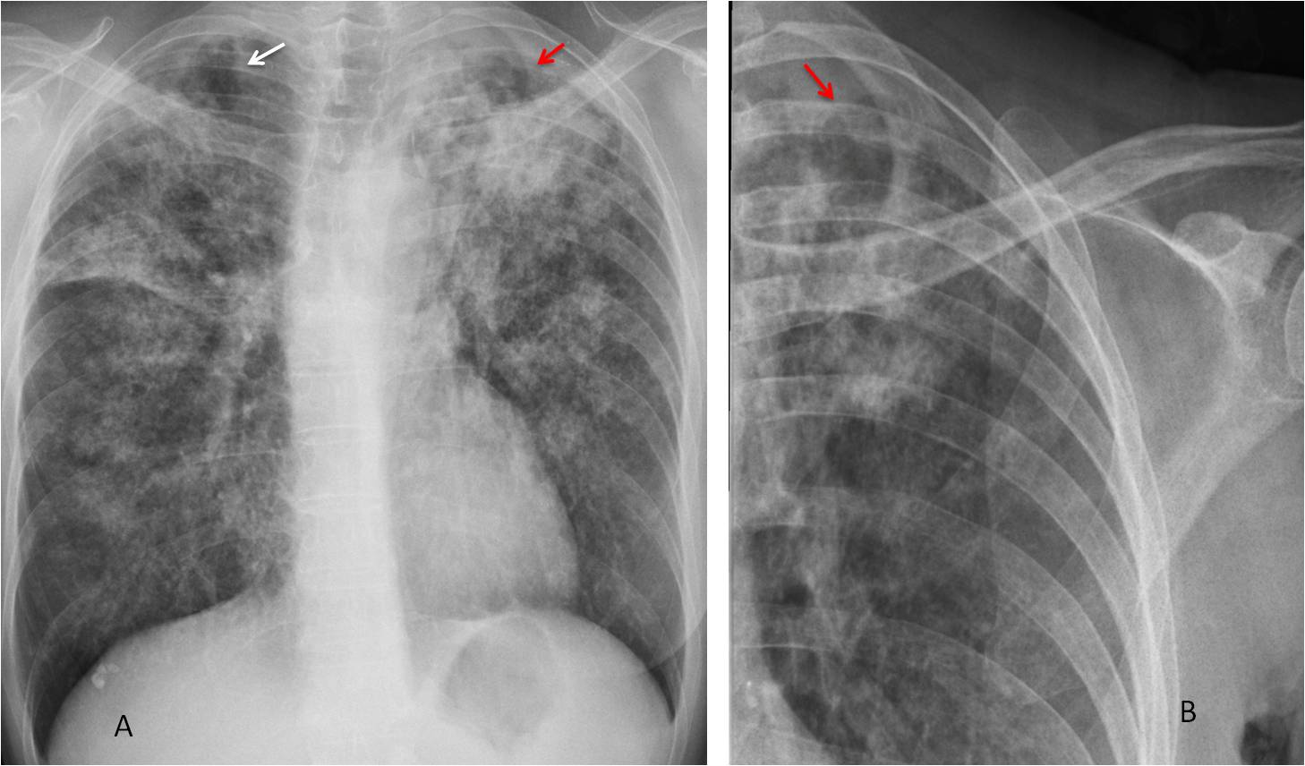
Fig. 1
Once cavitation is suspected, pulmonary TB is the diagnosis of choice, suggesting also that the nodule in the vocal cord may be a tuberculous granuloma. CT confirms the air space disease accompanied by cavities (Fig 2). BK was found in the sputum. Surgery was suspended. The pulmonary infiltrates and the lesion in the cord improved with treatment.
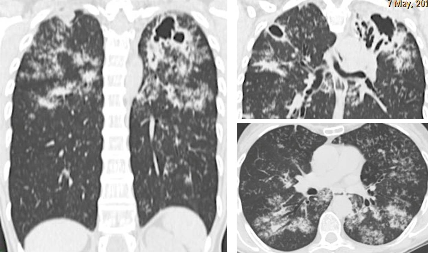
Fig. 2
Diagnosis: cavitated pulmonary TB with TB granuloma in vocal cord
Congratulations to Giovanni Battaglia, who was the first to suggest the right diagnosis.
This case emphasises several teaching points:
1. Paucisymptomatic TB is not rare, especially in middle class, well-nourished individuals.
2. Discovering cavitation is important to suspect TB
3. Remember the association of laryngeal disease with active pulmonary TB. I have seen several cases.