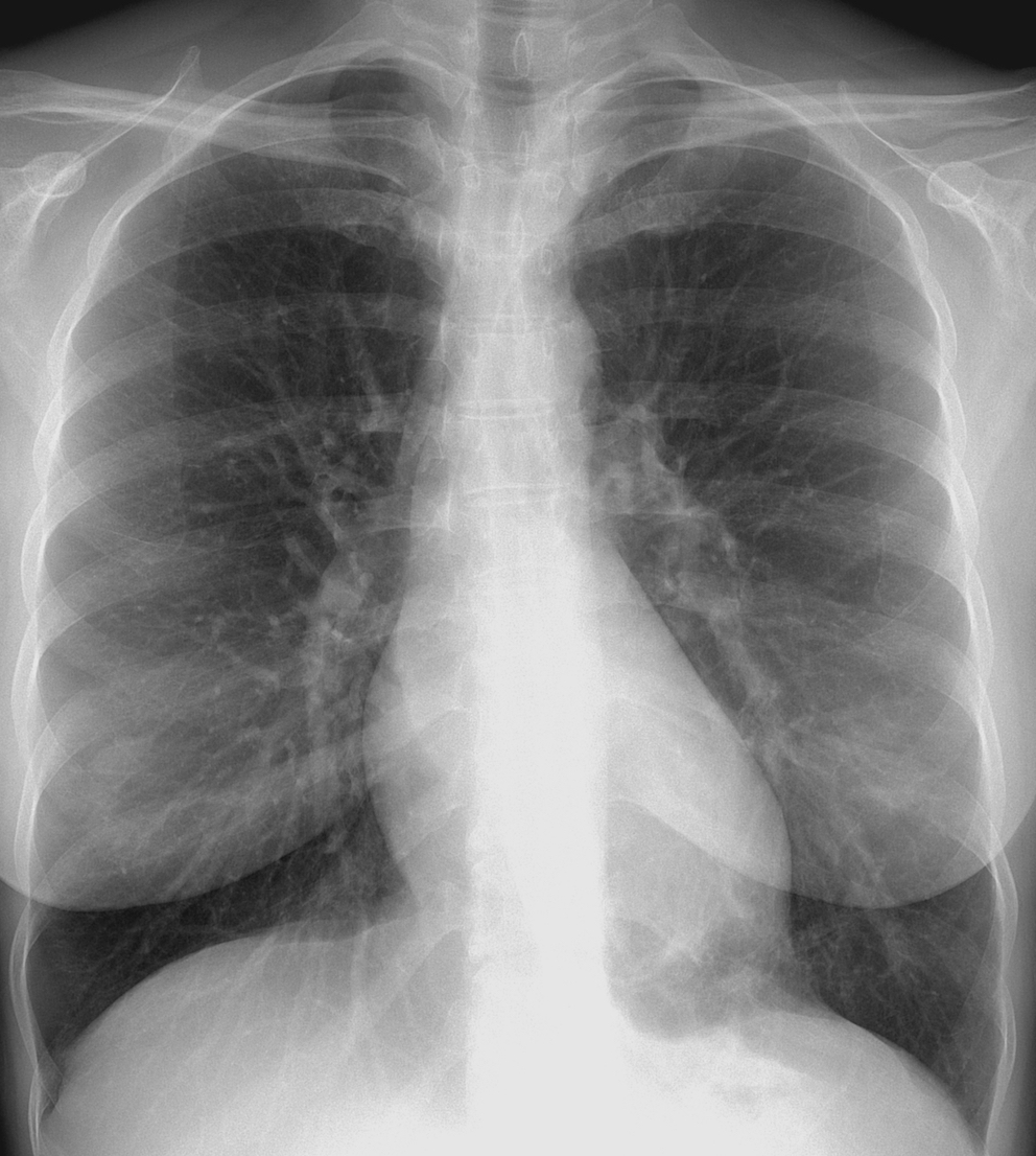
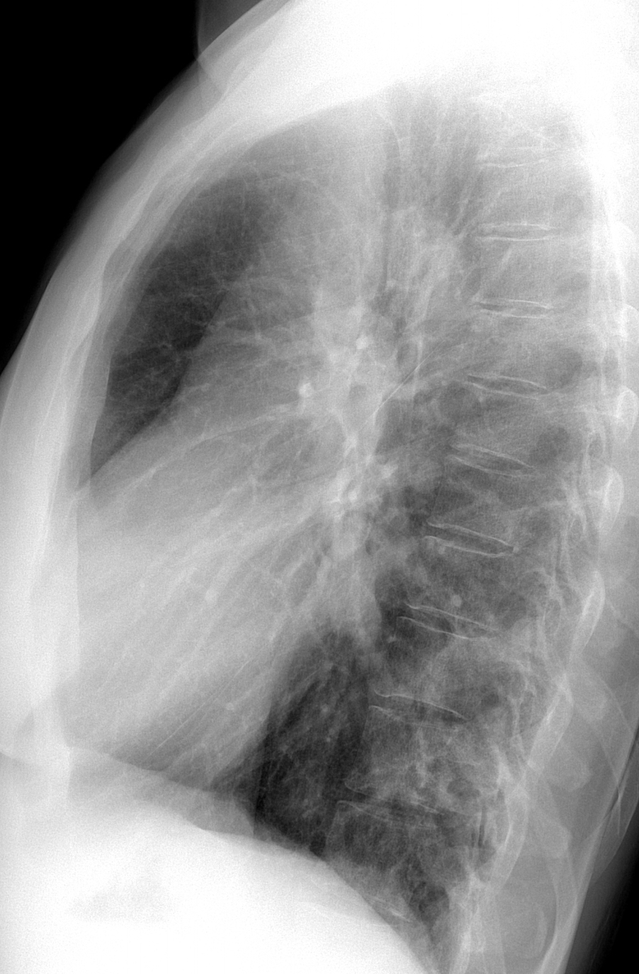
Today we are showing a case that our good friend Carles Vilá showed us three weeks ago. Images belong to a 44-year-old woman admitted with renal colic. What do you see?
Check the images below, leave your thoughts in the comments section and come back on Friday for the answer.
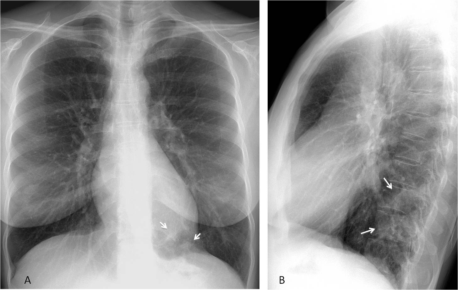
Findings: the lateral view shows an increased opacity at the level of the T10-T11 vertebral bodies (B, arrows), which is visible as ill-defined opacities at the left lung base in the PA view (A, arrows). They are not specific for diagnostic purposes.
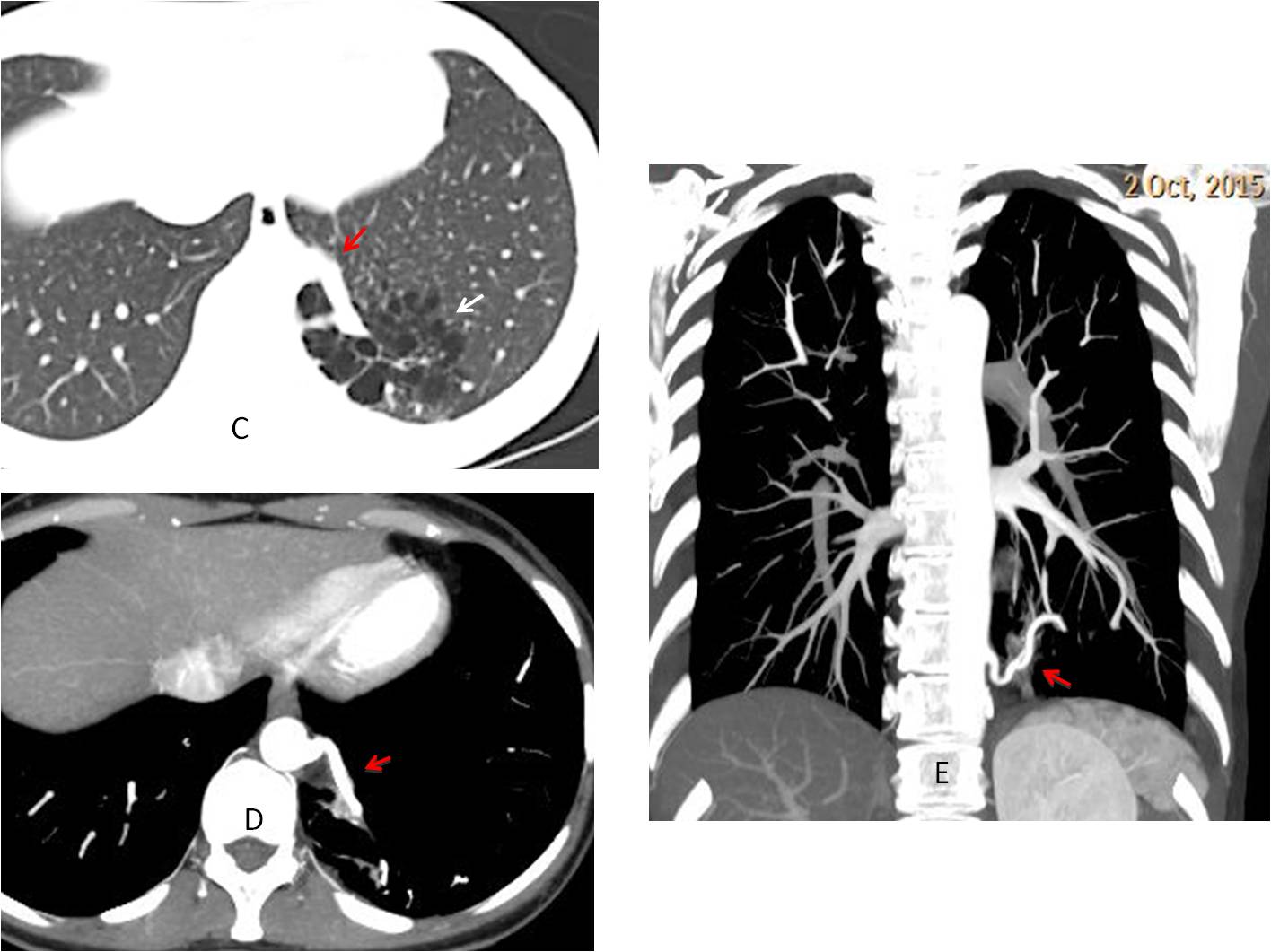
Unenhanced abdominal CT for the study of renal colic discovered a multicystic lesion in the left lung (C, arrow) with a possible abnormal vessel arising from the abdominal aorta (C, red arrow), confirmed with enhanced chest CT (D-E, red arrows).
When questioned, the patient denied any episode of previous pulmonary infection.
Final diagnosis: cystic sequestration of the lung
Congratulations to Ryan, who was the first to observe the abnormality
I am showing this case because it is a nice complement to case 121, in which the increased opacity of the vertebral bodies was due to spinal disease. In the present case, the opacity is also obvious and due to superimposed lung pathology.
Teaching point: Remember that congenital lesions are not rare in the adult and are seen more often than expected.