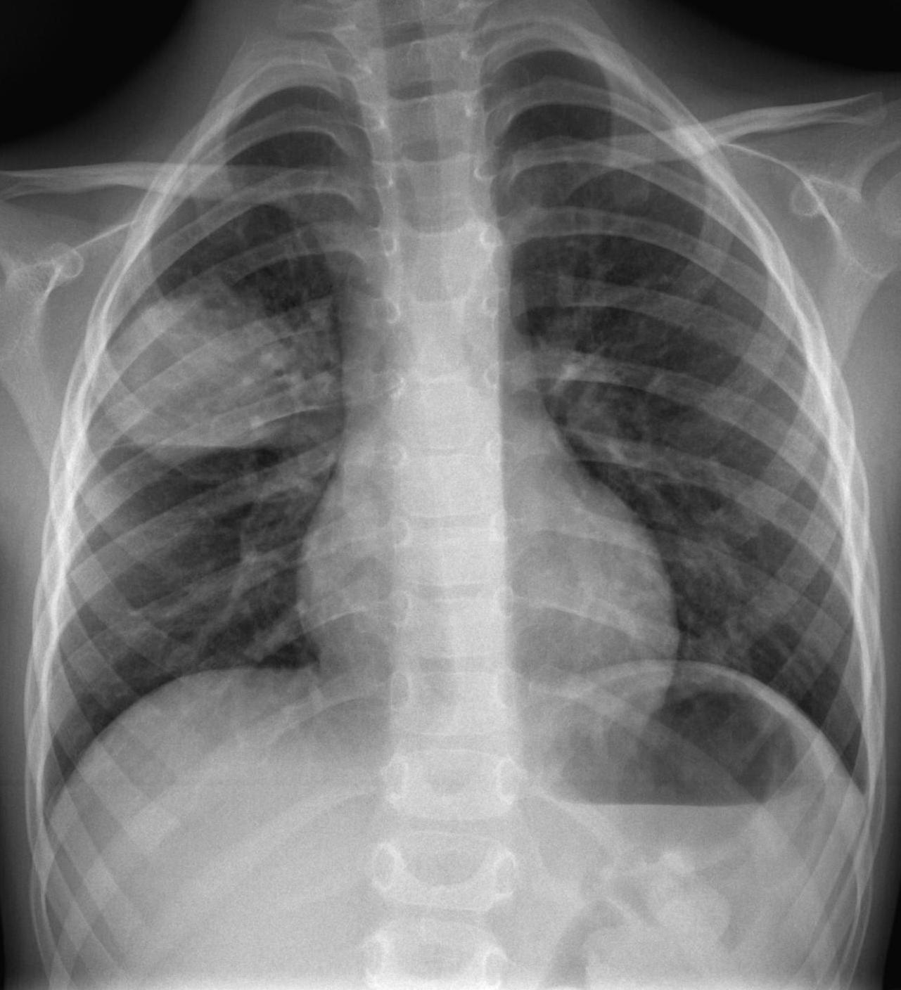
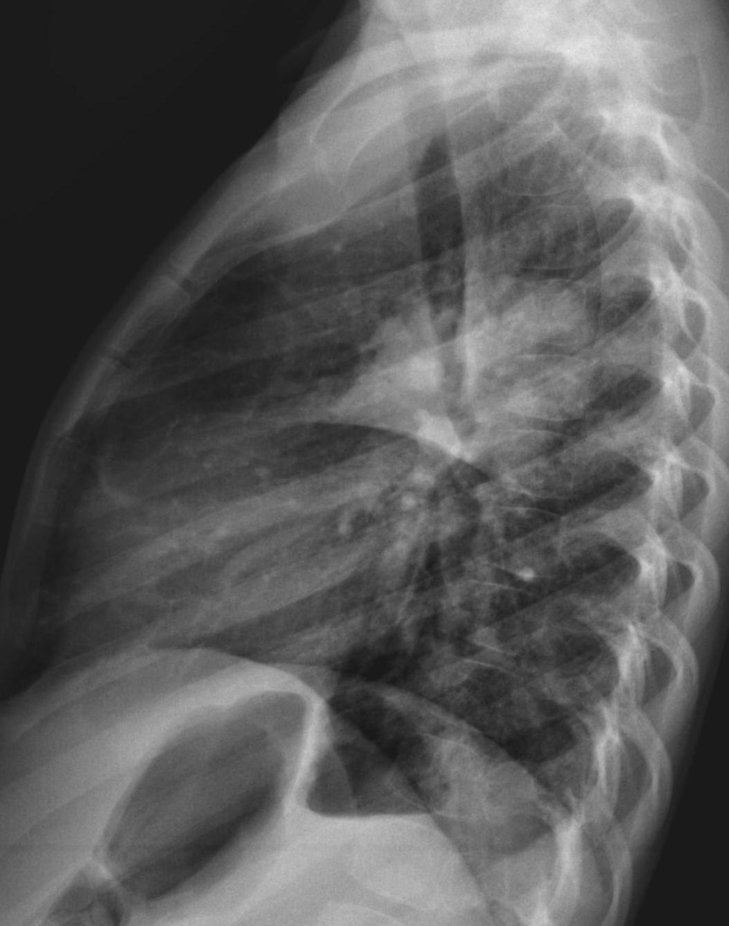
To mark the International Day of Radiology, which this year is focusing on paediatric imaging, we are presenting chest radiographs of a 5-year-old boy with an abdominal mass and mild fever. A Wilm’s tumour was suspected. Check the images below, leave your thoughts in the comments section and come back for the answer on Friday.
Diagnosis.
1. Pleural tumour in fissure
2. Pneumonia
3. Metastases
4. None of the above
Findings: chest radiographs show a rounded pulmonary lesion in the RUL. The indistinct borders, except where contacting with the minor fissure (A-B, red arrows), exclude the diagnosis of tumour or intracisural lesion.
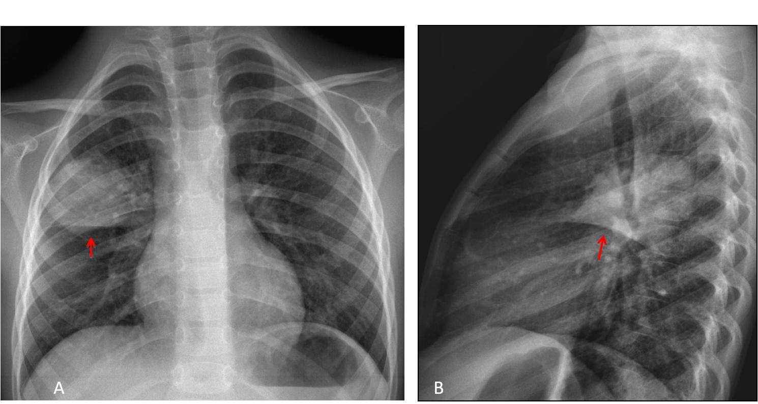
The most likely diagnosis is pneumonia, especially in the presence of fever. Chest radiographs taken ten days later (C-D) confirm the disappearance of the lesion.
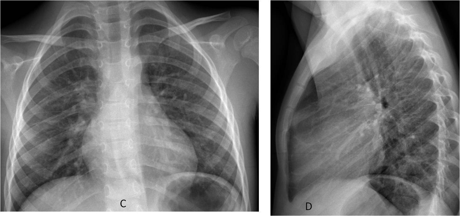
Final diagnosis: round pneumonia
My apologies if the diagnosis was too easy, but I didn’t have the heart to give this child (any child) a malignant disease.
Congratulations to all of you who made the right diagnosis, led by Murzin, who was an early starter.
Teaching point: when faced with a round pulmonary lesion in a child, always consider round pneumonia as the first possibility.