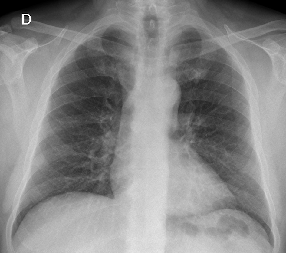
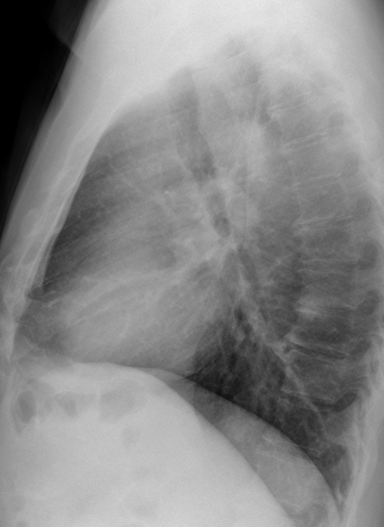
Today we are showing radiographs of a 59-year-old man with chest pain. What do you see?
Check the images below, leave your thoughts in the comments section, and come back on Friday for the answer.
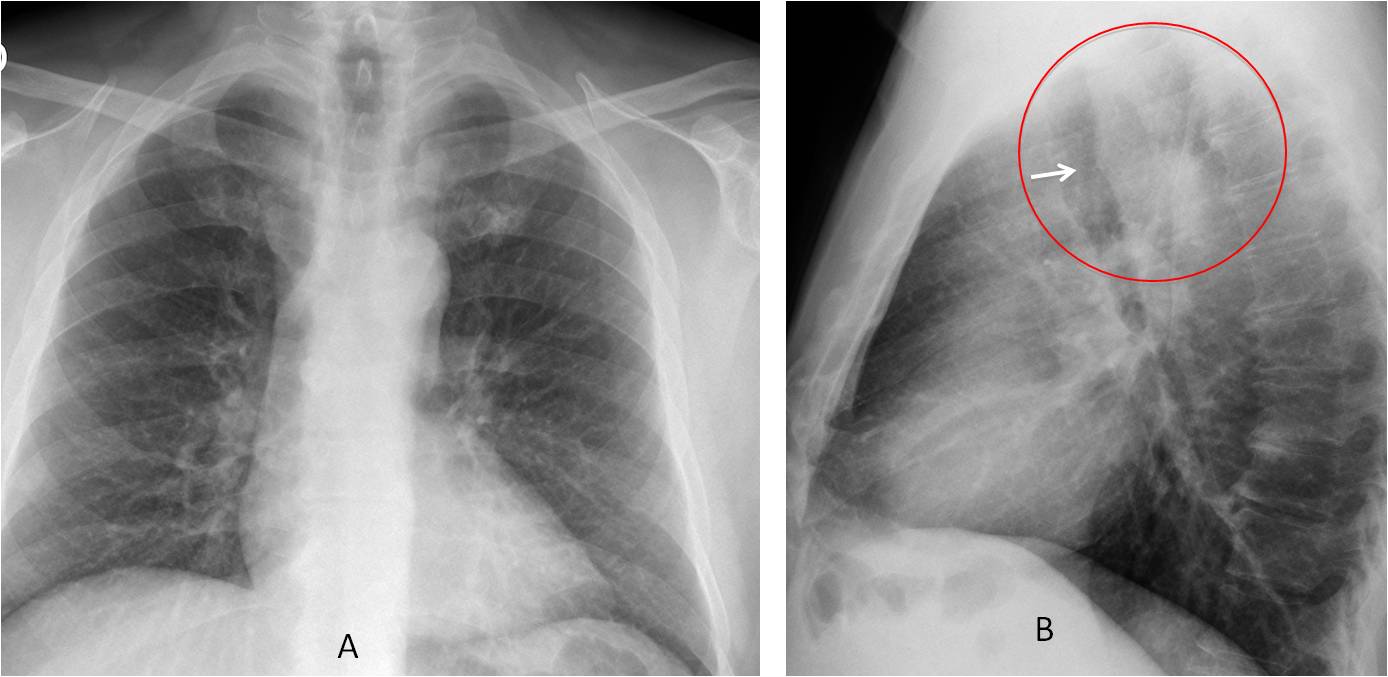
Findings: the PA radiograph (A) is unremarkable. The lateral view shows a slight protrusion of the posterior tracheal wall (B, arrow), accompanied by increased opacity of the retrotracheal space (B, circle) with disappearance of the posterior paratracheal line. These findings are highly suspicious of a posterior mass (see
Dr Pepe’s Diploma case no. 72), most likely arising from the oesophagus. Vascular malformation is less likely in the presence of pain.
Coronal CT shows a retrotracheal mass (C, arrow). Sagittal CT reconstruction mimics the findings in the lateral view (D-E, circle).
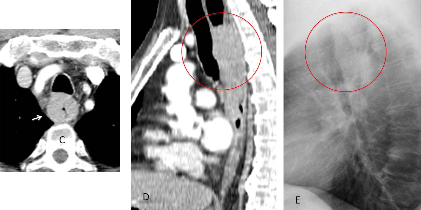
Final diagnosis: carcinoma of oesophagus
I showed this case to emphasise the importance of the trachea and retrotracheal space in the lateral view. All of you who mentioned abnormalities in this area will get a Gold Medal, with special mention to Ivan.
Teaching point: always remember to look at the trachea in the lateral view. It may offer important information, as in the present case.