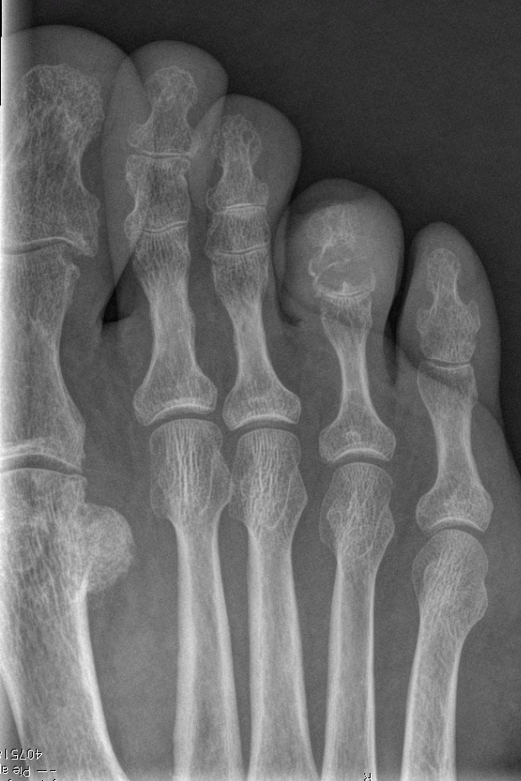
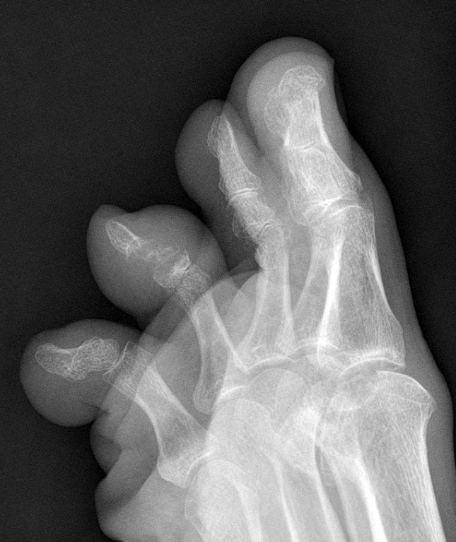
You may wonder what a foot is doing in a chest blog. The answer is obvious: because the foot belongs to a chest radiologist. These radiographs are of my own foot, where I noticed a painless swelling in the 4th right toe, present for about one year. Check the images below, leave me your thoughts and diagnosis in the comments, and come back on Friday for the answer.
Findings: AP and oblique views of the left foot show a lytic lesion affecting the distal joint of the fourth toe, extending into the distal and middle phalanges. The borders of the osseous lesions are sclerotic, indicating a slow-growing process (A-B, arrows). There is an accompanying soft-tissue mass (B, red arrow). The mass does not enhance after gadolinium injection in the MRI study.
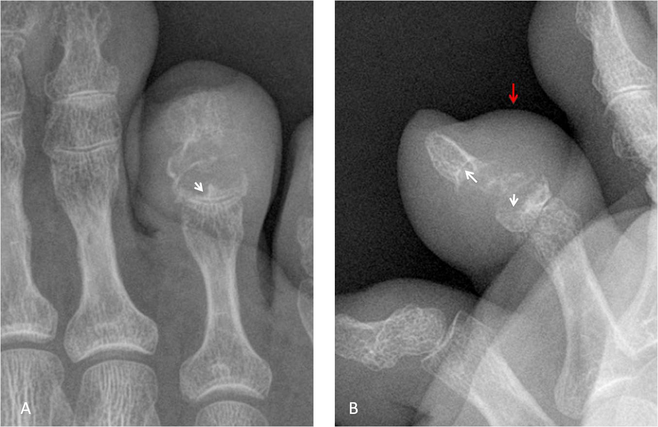
The plain film appearance of the lesion is typical of the so-called lumpy-bumpy arthritis. The differential diagnoses include: gout, sarcoidosis, pigmented villonodular synovitis and amyloid, among others. Percutaneous biopsy confirmed the presence of uric acid crystals.
Final diagnosis: tophaceous gout.
Congratulations to Aloria, who was the first to suggest the diagnosis.
Teaching point: I am showing this case to emphasise the importance of knowing basic semiology. In retrospect, the lesion is a textbook example of lumpy-bumpy arthritis, of which gout is the most common etiology. My colleagues and I were too scared to think clearly. On the other hand, I am proud to have the same disease as my mentor, Dr. Benjamin Felson.