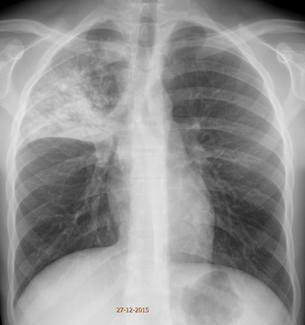
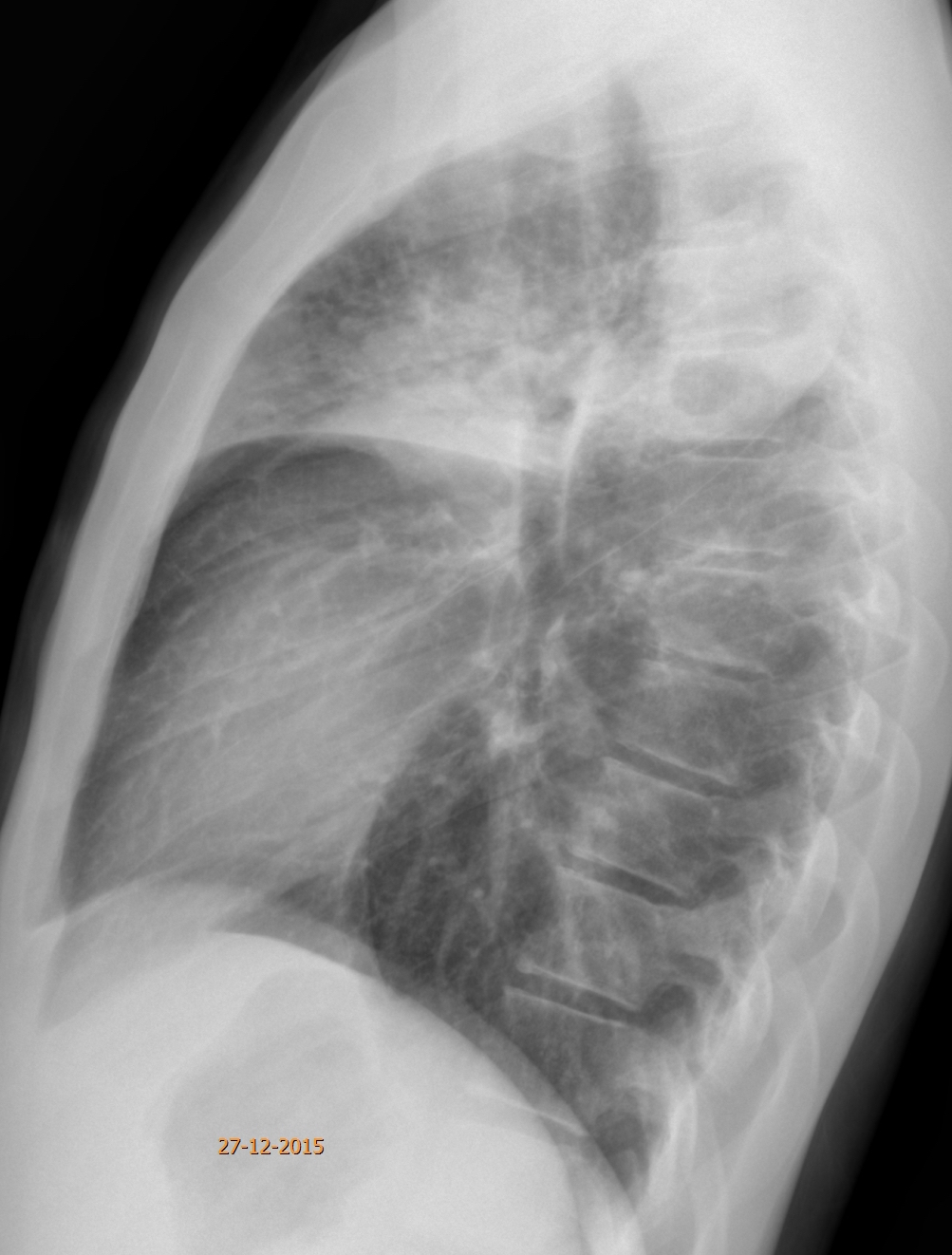
This week’s images belong to a 16-year-old boy with a cough and fever for the last two weeks.
Check the images below, leave your thoughts in the comments section, and come back on Friday for the answer.
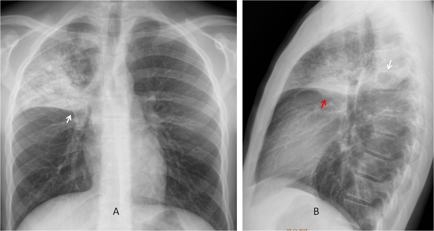
Findings: PA radiograph shows RUL disease with a suggestion on a Golden’s sign (A, arrow). There is an obvious cavity in the infiltrate in the lateral view (B, arrow). There is also a hint of a hilar mass or enlarged lymph nodes (B, red arrow).
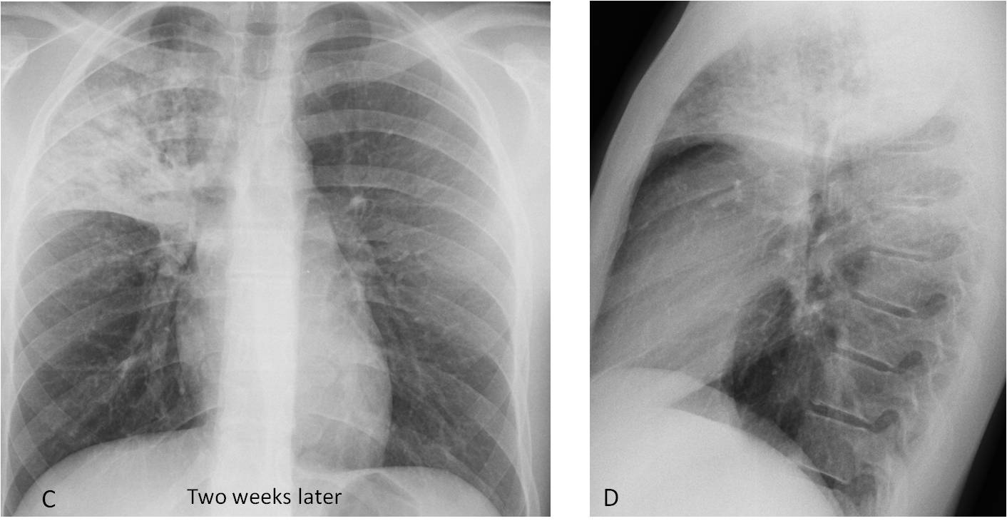
The patient was treated for pneumonia and two weeks later the appearance of the chest was practically unchanged (C-D).
Axial and coronal CT show multiple cavities within the RUL infiltrate (E-F, red arrows), and areas of tree-in-bud in the periphery (E, circle). Sputum yielded abundant Mycobacterium tuberculosis bacilli.
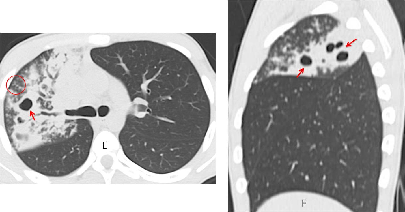
Final diagnosis: Acute lobar TB
Congratulations to all of you who suspected TB, led by Leila, who was the first to mention the diagnosis
Teaching point: Always look at the lateral view. The cavity was missed in the first examination, delaying the diagnosis.