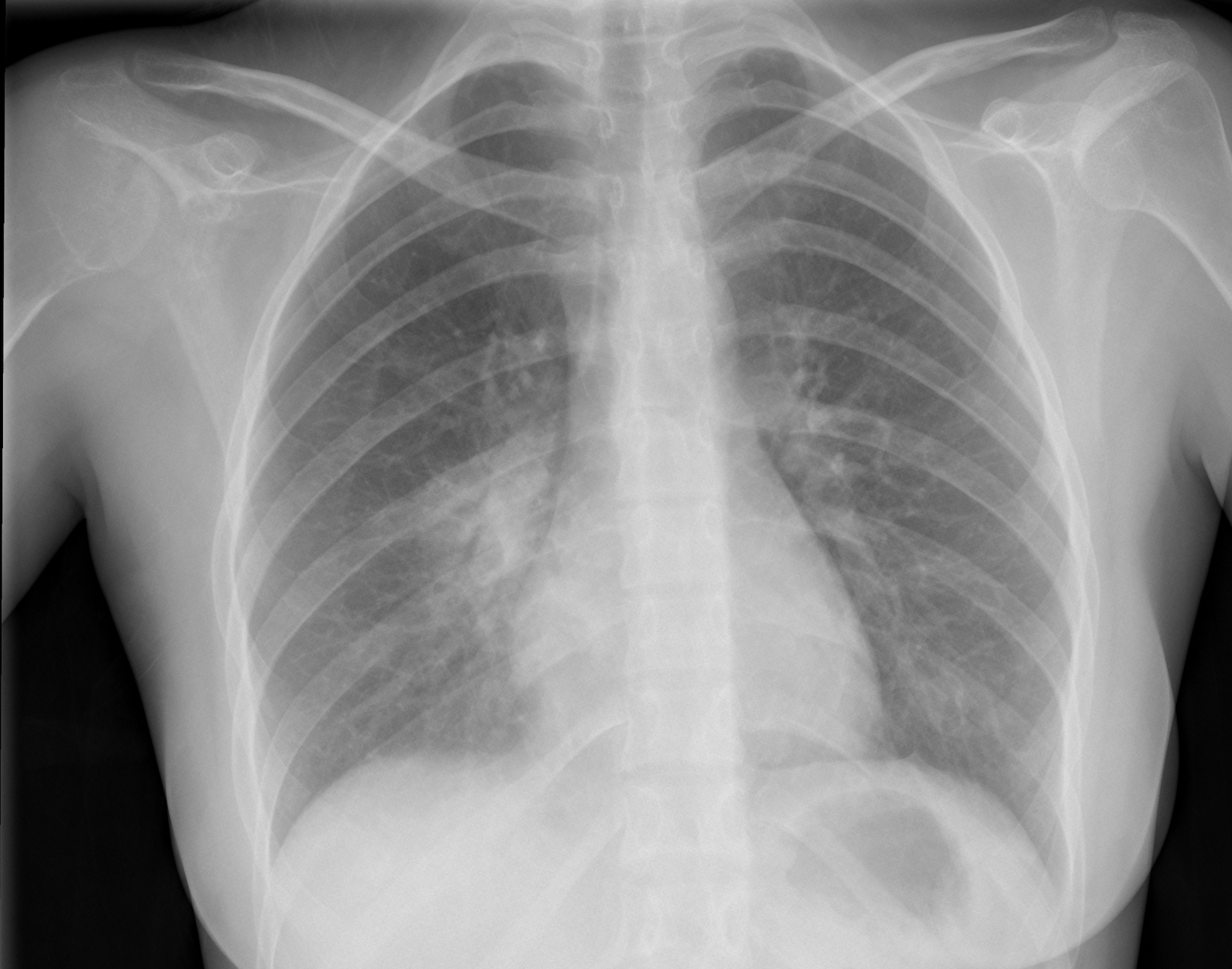
Today we are presenting a PA chest of a 28-year-old woman with fever and pain in the right hemithorax. What do you see? Check the images below, leave your thoughts in the comments section, and come back on Friday for the answer.
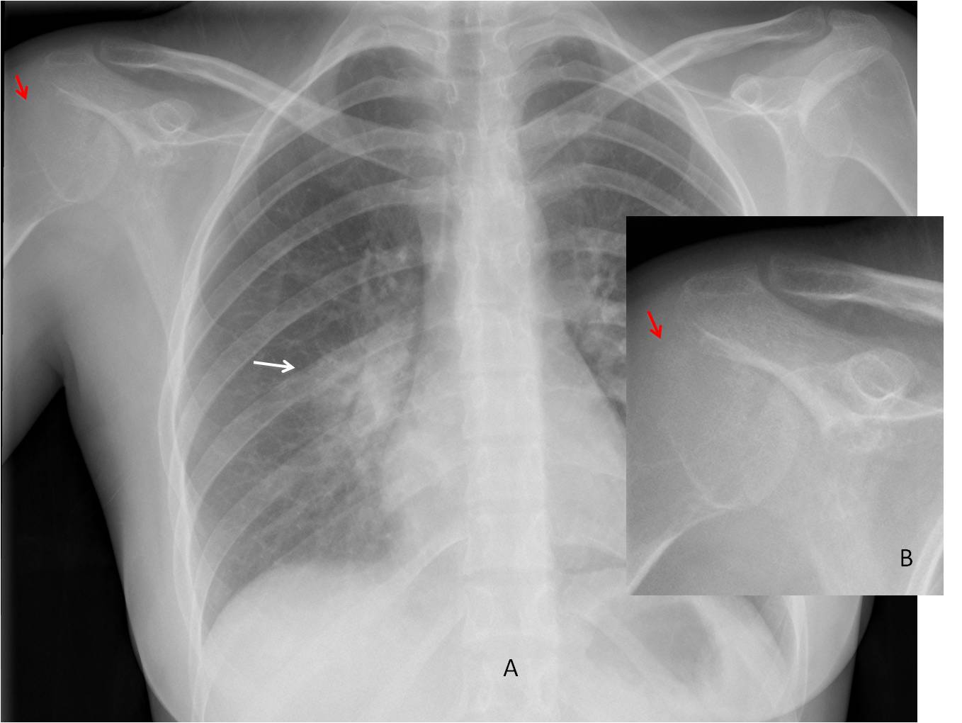
Findings: PA radiograph shows air-space disease around the right hilum and RLL (A, arrow). Given the age of the patient and the clinical symptoms, the most likely diagnosis is acute pneumonia. There is also loss of opacity and lack of visualisation of the outer cortex of the right humeral head (A, red arrow). These findings are more evident in the magnified view (B, red arrow) (compare with left shoulder, above).
PA radiograph of the right shoulder shows a slightly expansive lytic lesion of the humeral head with numerous bone ridges inside (C, arrow). CT confirms the findings (D, arrow).
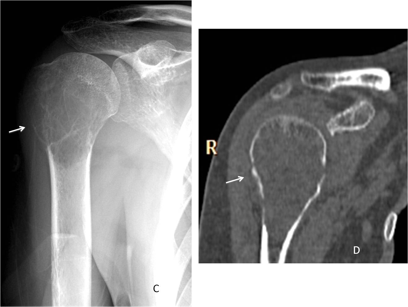
Diagnosis: Giant-cell tumour of humeral head
Congratulations to Murzin, who was the first to detect the humeral lesion.
Teaching point: remember to avoid satisfaction of search. It is important to look at the whole radiograph, because other pathologies may be found, as in this case