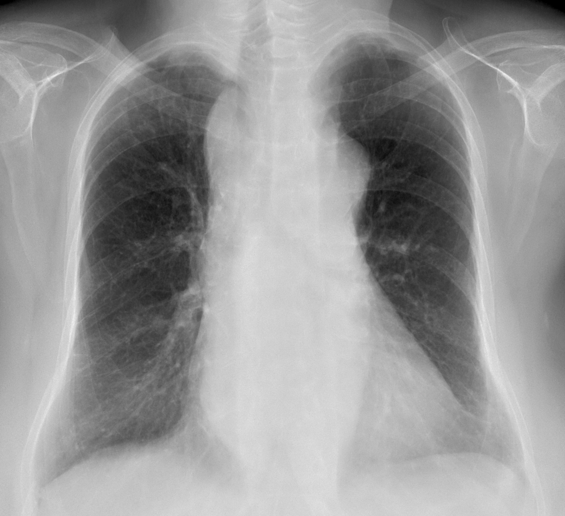
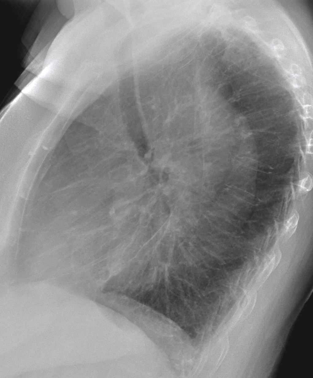
Considering that this is Holy Week, I want you to do penance. This week’s images belong to a 73-year-old woman with mild dyspnoea and vague chest complaints.
What do you see? Check the images below, leave your thoughts in the comments section and come back on Friday for the answer.
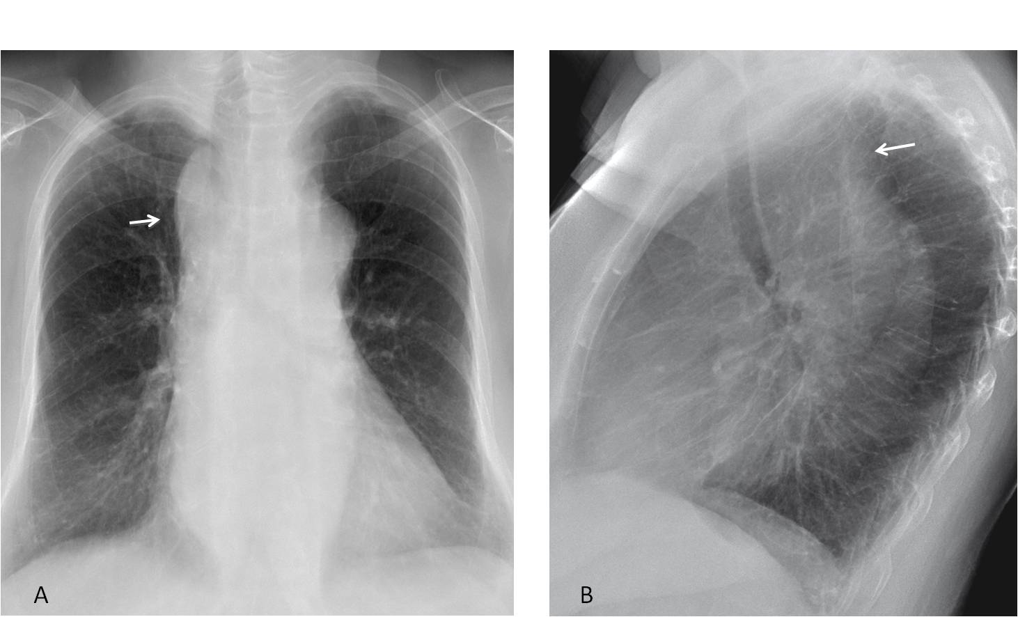
Findings: the PA radiograph shows widening of the right superior mediastinum (A, arrow), which in the lateral view is located behind the trachea (B, arrow). The initial impression is of an upper middle mediastinal mass. The first diagnoses that come to mind are a goiter or a dilated aorta.
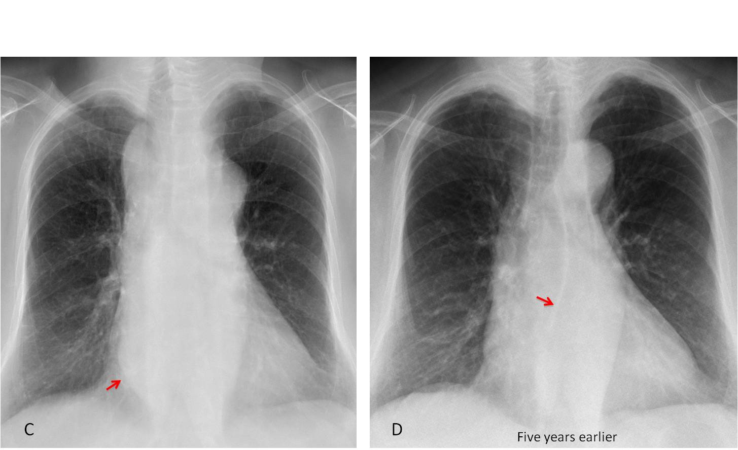
However, this is a typical case of satisfaction of search (again!). Most of you have missed the bulging of the azygo-oesophageal line (C, arrow), which is more evident compared with a film taken five years earlier (D, arrow). Therefore, we are dealing with a mass that extends along the middle mediastinum from top to bottom. The findings point to a dilated oesophagus or, more rarely, to a dilated aorta.
Oesophagogram was unremarkable. Coronal and sagittal CT shows a cystic tubular mass extending along the posterior wall of the oesophagus (E-F, arrows).
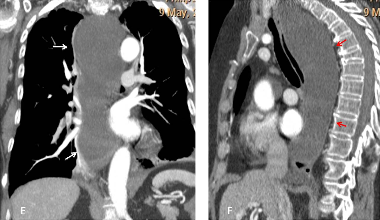
Final diagnosis: cystic lymphangioma of mediastinum.
This is a difficult case and I didn’t expect you to make the diagnosis. But I believe that you should have noticed the bulging of the azygo-oesophageal line and suggested a dilated oesophagus, which is the most sensible diagnosis.
Kudos goes to Bujar, who was the one to get closer to the truth.
Teaching point: Failing to look beyond the obvious is the cause of about 20% of misdiagnosis.