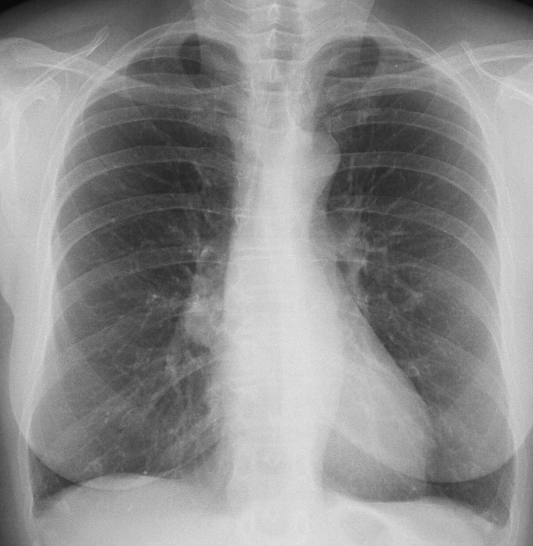
Today I am showing the second case of the “five easy pieces”. It is a PA radiograph of a 51-year-old woman for pre-op surgery of umbilical hernia. What do you see?
Check the image below, leave me your thoughts in the comments section, and come back on Friday for the answer.
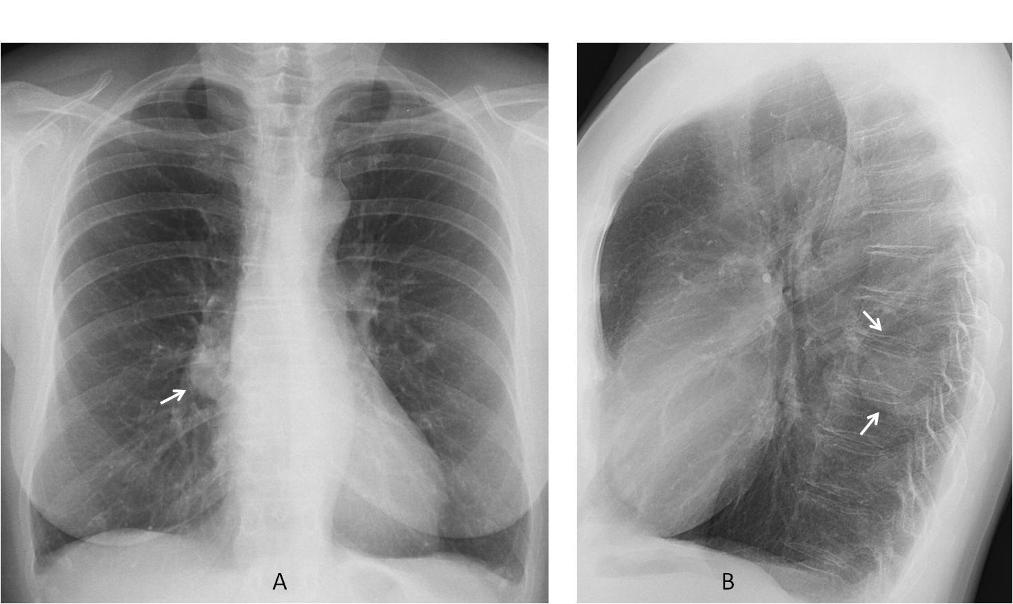
Findings: PA chest radiograph shows increased opacity of the lower right hilum (A, arrow). Lateral view was taken and demonstrated that the opacity was located posteriorly (B, arrow) and had features of an extrapulmonary lesion.
Enhanced CT confirms a solid posterior mediastinal mass (C-E, arrows). The most likely diagnosis is a neurogenic tumour. At the time of writing, surgery is still pending.
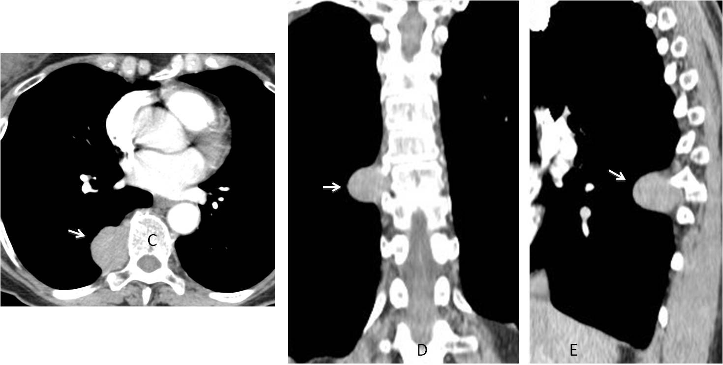
Final diagnosis: posterior mediastinal mass simulating an enlarged hilum.
Congratulations to all of you (many) who saw the abnormality, with special mention to Olena, who gave the first answer.
Teaching point: the most common cause of prominent hilum is lymph nodes or an enlarged pulmonary artery. Occasionally it may be simulated by superimposition of a mass located in front of or behind the hilum, as in this case.