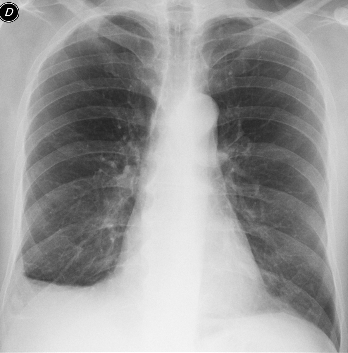
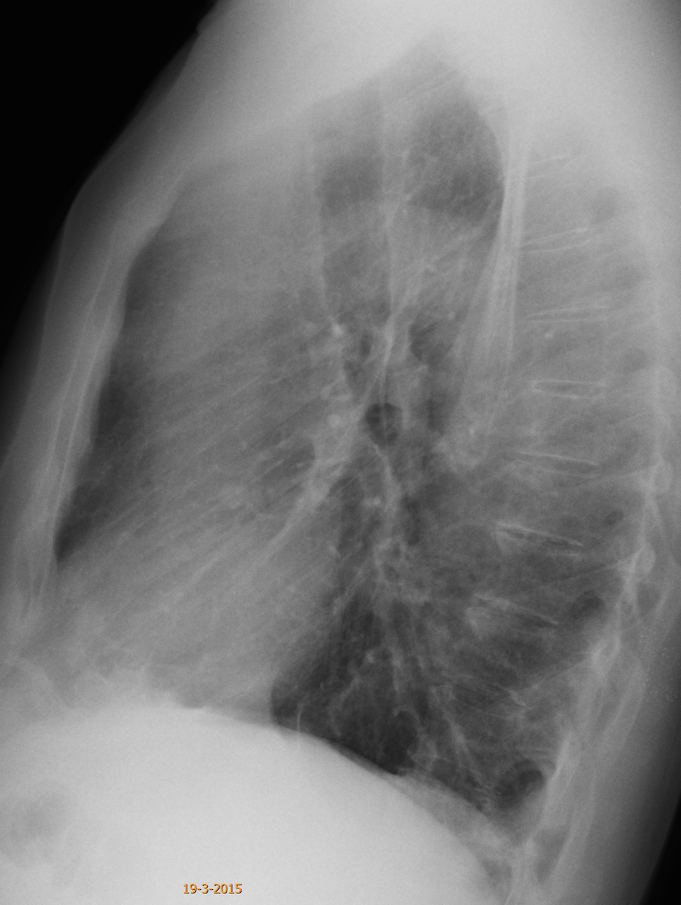
Today I am presenting the third case of the “five easy pieces”. Images belong to a 63-year-old man operated on for carcinoma of the colon five years ago. Previous annual CTs were read as normal. What do you see?
Check out the images below, leave your thoughts in the comments section and come back on Friday for the answer.
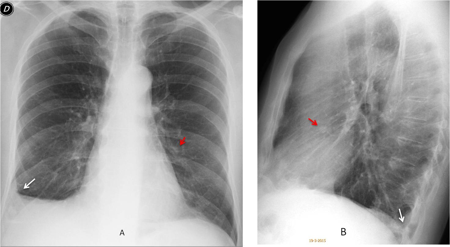
Findings: the most important finding is a small pulmonary nodule seen in the lateral view (B, red arrow) and barely visible in the PA (A, red arrow). Considering that the patient has a primary tumour, metastases should be suspected. There is also obvious blunting of the right costophrenic angle (A, arrow), which looks like scarring because of the straight diaphragm and the absence of blunting on the posterior sulcus (B, arrow).
Axial CT confirms the presence of the nodule (C, arrow). Free fluid was not present. Review of previous studies shows that the nodule was overlooked in previous CTs, taken in 2014 and 2010 (D-E, arrows). Surgery confirmed metastases.
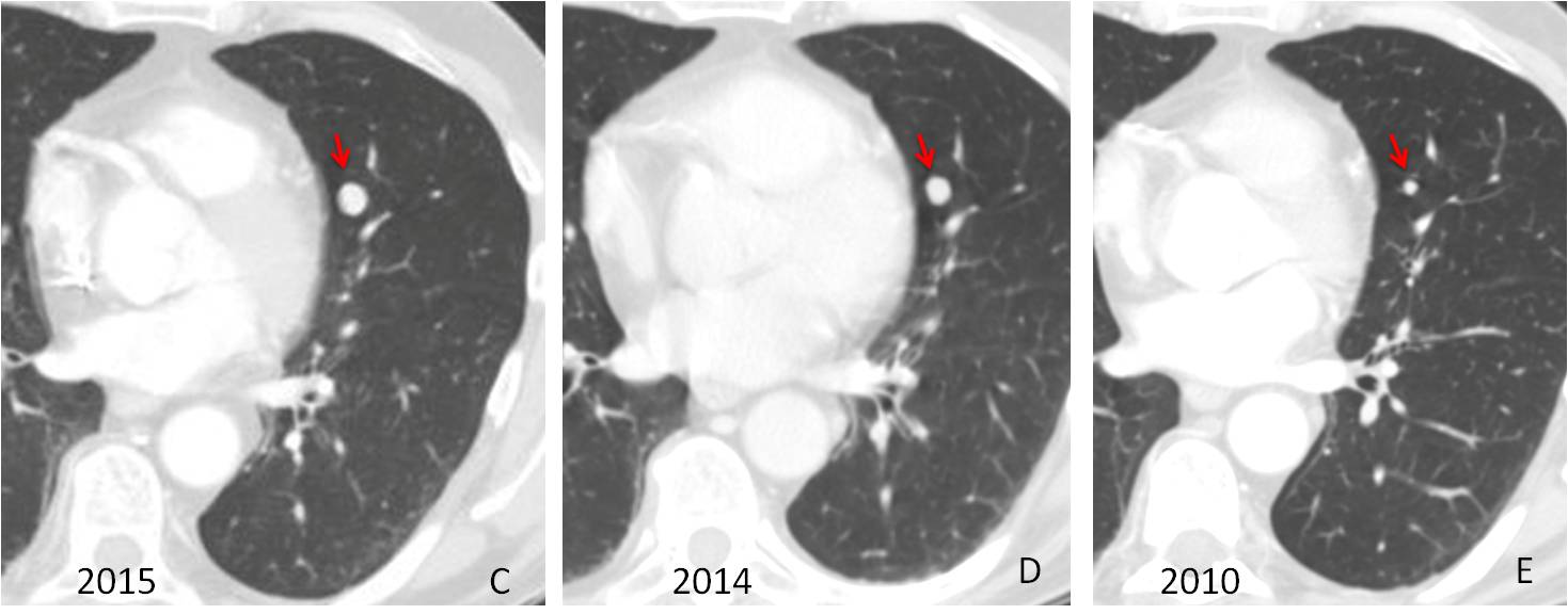
Final diagnosis: solitary metastases, overlooked at previous CTs and discovered in the chest radiograph.
Congratulations to all of you who saw the metastases, led by Olena, who was the first to make the diagnosis.
Teaching point: this case is a demonstration of the value of the lateral film, demonstrating a metastatic nodule that was overlooked at CT.