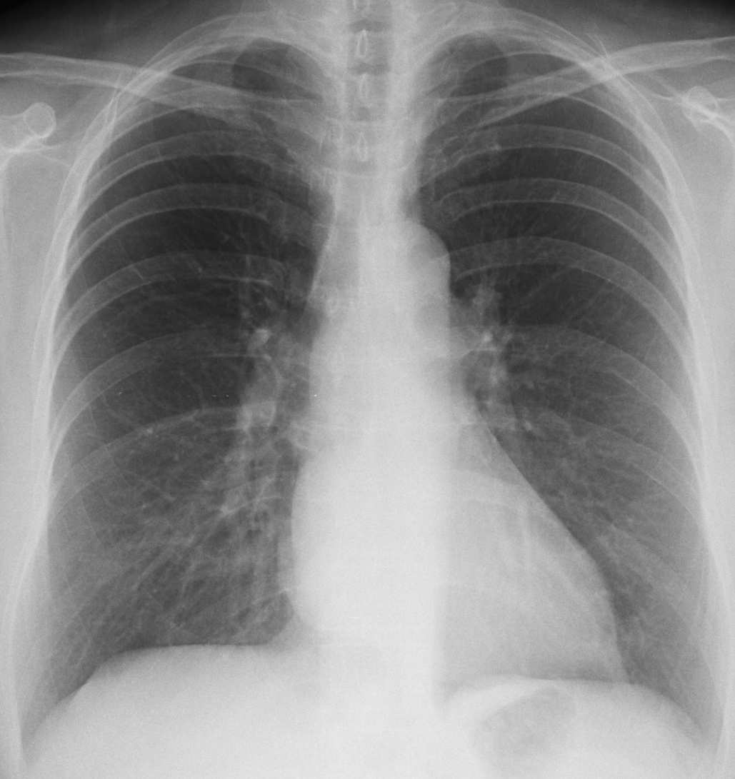
This is the fourth case of the “five easy pieces”. Pre-op chest radiograph for lumbar hernia of a 54-year-old woman.
What do you see? Check the image below, leave your thoughts in the comments section and come back on Friday for the answer.
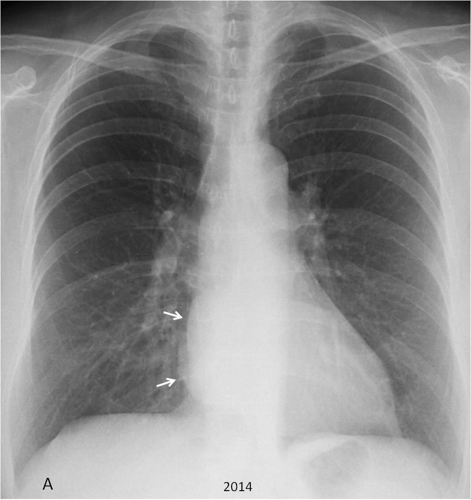
Findings: PA chest taken in 2014 shows a double contour of the right heart border (A, arrows), which suggests a retro cardiac mass, either in the middle or posterior mediastinum.
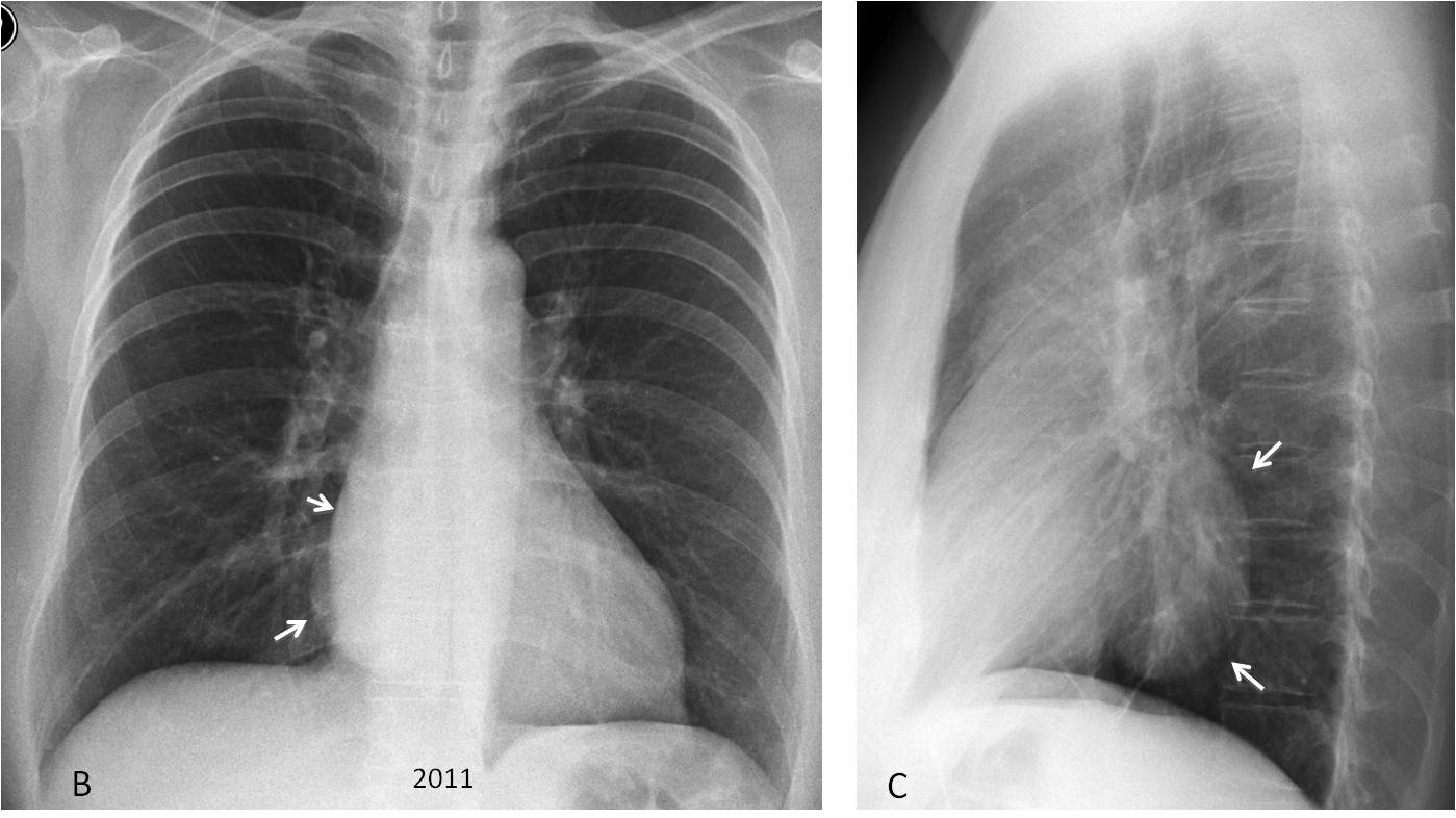
Review of previous films taken in 2011 confirms identical findings in the PA view (B, arrows), which in the lateral view correspond to an ovoid middle mediastinal mass (C, arrows).
A recent study from 2016 shows that the mass is unchanged ((D-E, arrows). Because of this lack of change, a CT was not done.
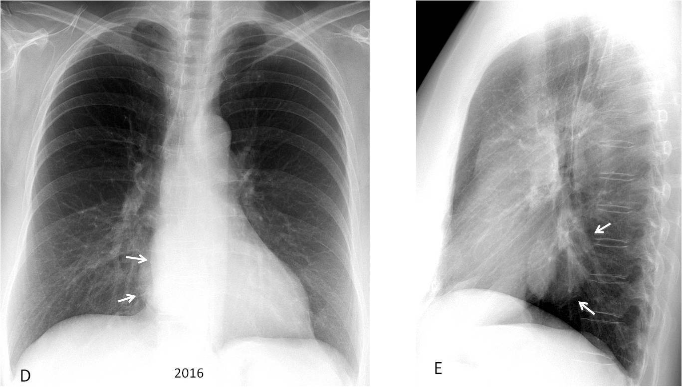
Final diagnosis: probable mediastinal duplication cyst (unproven).
Congratulations to Eugeny, who was the first to notice the abnormality, unlike the radiologist who saw the chest in 2014 and read it as normal.
Teaching point: this case emphasises the importance of taking a lateral view. And looking at previous films, that in this patient were available in the PACs station.