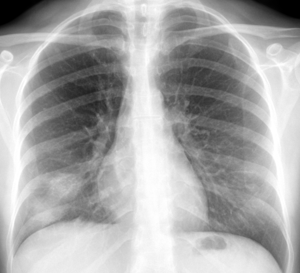
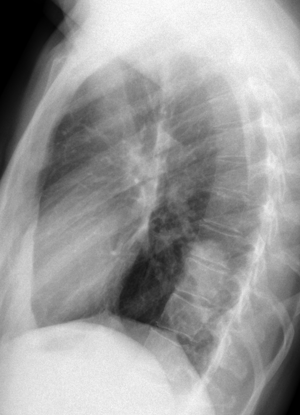
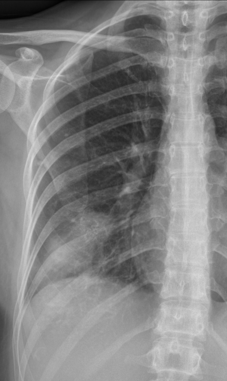
Dr. Pepe is back and he refuses to show easy cases. He has forced me to present the following images which belong to a 38-year-old woman with right chest pain. Check the three images below, leave us your thoughts in the comments section and come back on Friday for the answer.
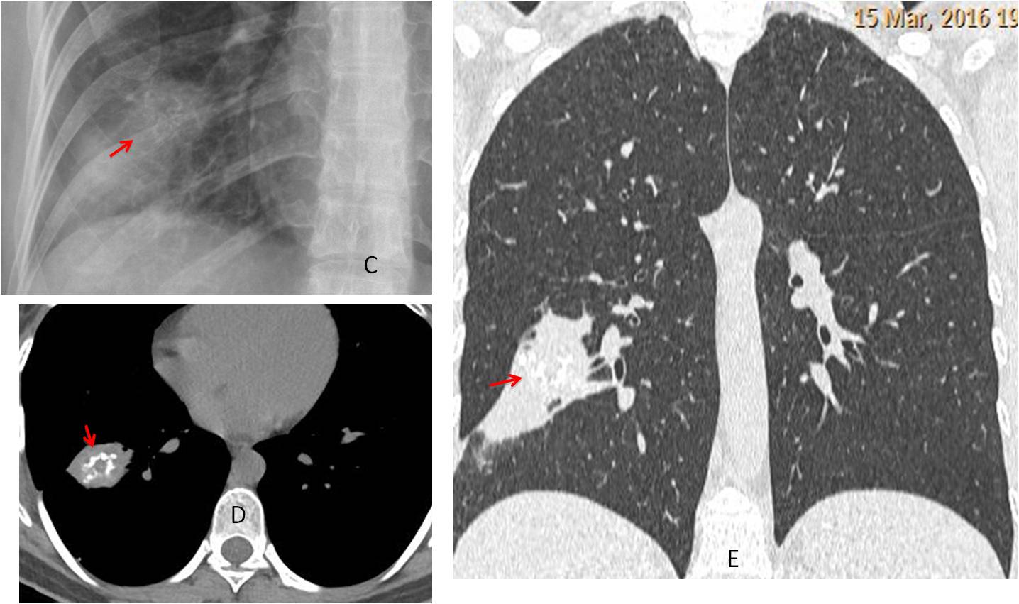
Findings: chest radiographs show an ovoid ill-defined opacity in the RLL (A-B, arrows). There are calcifications within it, better seen in the view of the ribs (C, red arrow).
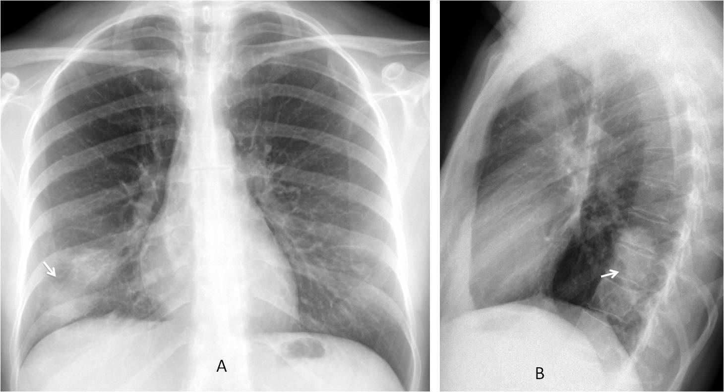
The presence of visible calcifications excludes the diagnosis of carcinoma. The ill-defined contour and the oblong shape go against hamartoma. Chronic TB cannot be excluded, but I don’t like it.
Axial and coronal unenhanced CT confirms the pulmonary lesion adjacent to the major fissure. Calcifications are clearly visible (D-E, red arrows).
With the information provided by the chest radiograph we can assume that the lesion is chronic (calcium present), affects the parenchyma and does not give significant symptoms. A lesion that fits this description is an inflammatory pseudotumour, which was confirmed by lung biopsy.
An interest fact is that yesterday I searched in Google: “localized calcified chronic pulmonary infiltrate in right lower lobe” and got an article about unusual lung tumors in which the pseudotumor was mentioned (RadioGraphics 2002; 22:601-619).
Final diagnosis: inflammatory myofibroblastic tumour a.k.a. inflammatory pseudotumour.
Congratulations to all of you who fought bravely against insurmountable odds. I saw this case two months ago (see the date in the CT) and thought that you might appreciate it.
Teaching point: although this case is tough, an accurate interpretation of the findings leads to a short differential diagnosis that includes the final answer.