Dear Friends,
Welcome back! To start the academic year I would like to show a case seen by Dr. Pepe while I was vacationing in Minorca.
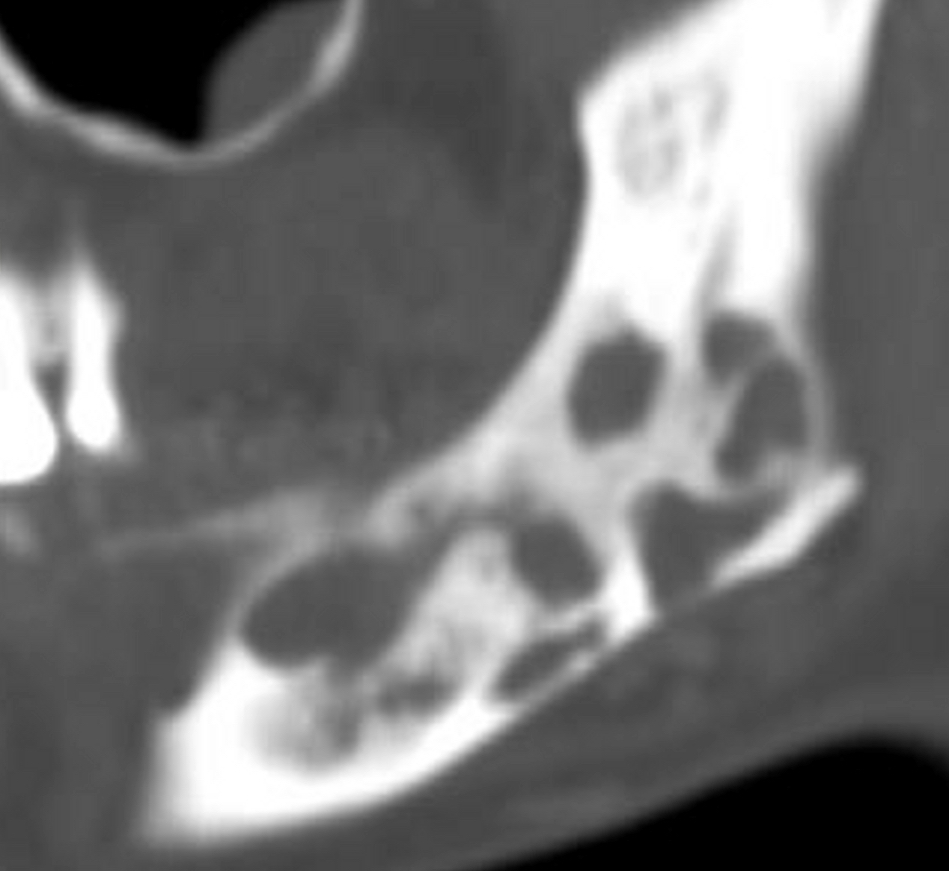
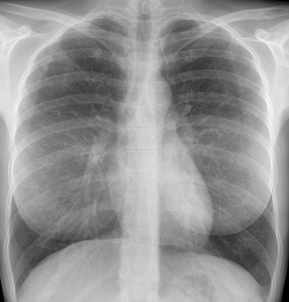
Radiographs belong to a 35-year-old woman with pain in the mandible for the last two years. CT of the mandible before biopsy and pre-op PA chest radiograph are shown. After seeing both, Dr Pepe suggested a diagnosis. What do you think?
Check the images below, leave your thoughts in the comments section, and come back on Friday for the answer.
CT mandible
PA chest
Findings: CT of the jaw (A) shows multiple lytic lesions, initially suspected to be a local tumor (adamantinoma). Pre- biopsy PA chest shows several small nodules in the upper lungs (B, circles).
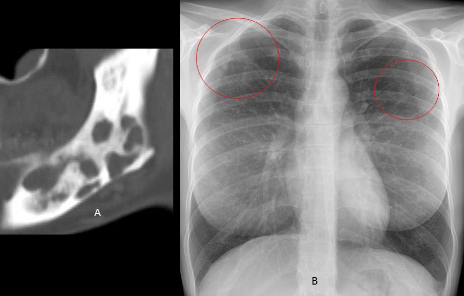
The combination of lytic bone lesions and nodules in the lung raised the suspicion of Langerhans -cell histiocytosis. The referring physician was contacted and an immediate CT was performed, showing numerous small lung nodules, the majority of them with cavitation (C, D). This appearance is highly suspicious of histiocytosis. Diagnosis was confirmed with bone biopsy. Referring physician was very impressed.
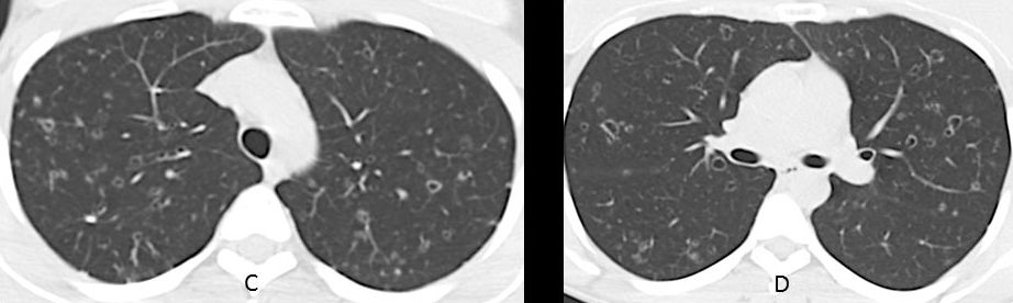
Final diagnosis: Langerhans-cell histyocitosis of lung and jaw.
Congratulations to GH and UFTM radiogroup for their early diagnosis
Teaching point: always look carefully at pre-op chest films. If interpreted correctly, they may facilitate the diagnosis.


