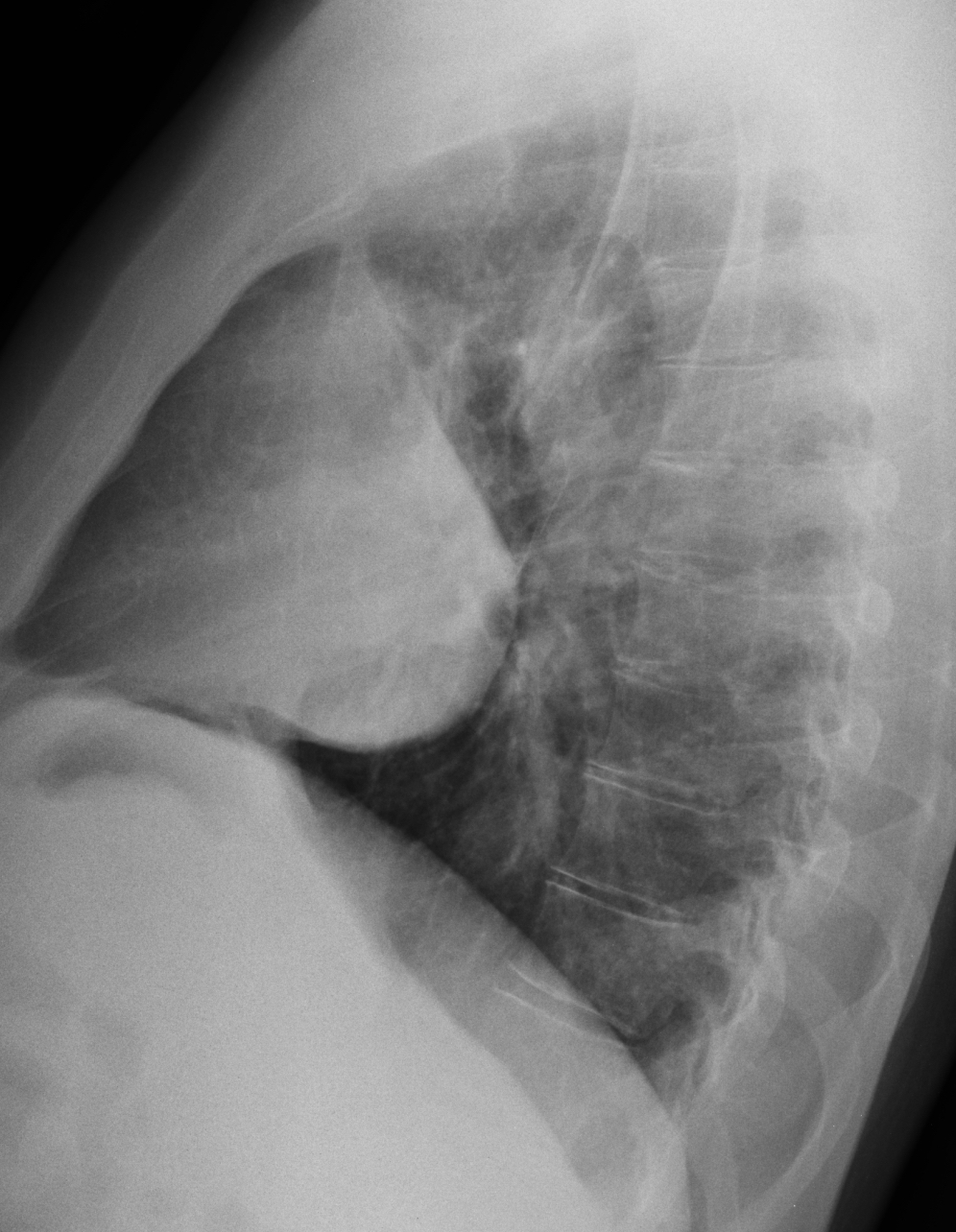
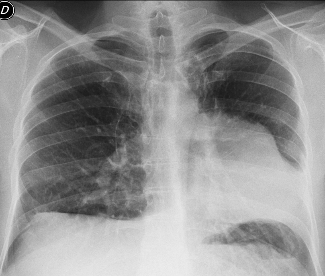
Today we are presenting pre-op radiographs for inguinal hernia in a 37-year-old-man. Check the images below, leave your thoughts in the comments section, and come back on Friday for the answer.
1. Pulmonary arterial hypertension
2. Thymoma
3. Pericardial cyst
4. None of the above
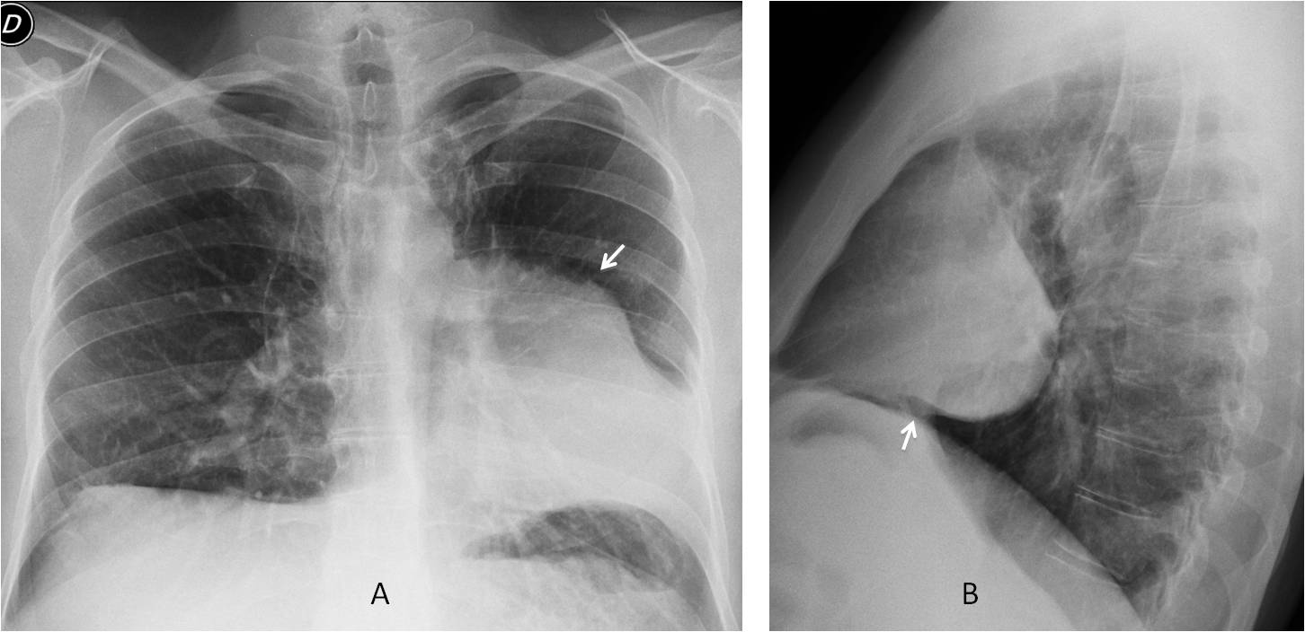
Findings: the PA radiograph shows an unusual shape of the heart, which is displaced towards the left and has a bumpy left border (A, arrow). The lateral view gives the clue to the diagnosis, by showing interposed lung between the diaphragm and the heart (B, arrow). In an asymptomatic patient, this sign is highly suspicious of absent pericardium.
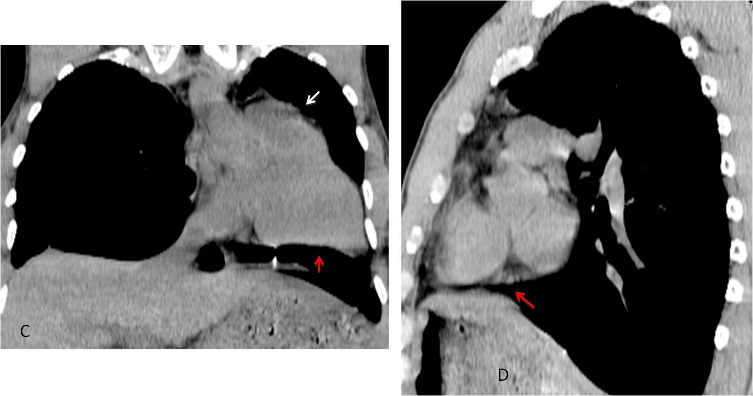
Unenhanced coronal and sagittal CT show lung under the heart in both views (C-D, red arrows). The lump in the left heart border corresponds to the pulmonary artery (C, arrow).
A chest radiograph taken five years earlier (not shown) showed identical appearance.
Final diagnosis: congenital absence of pericardium.
Congratulations to Diogo, who was the first to make the diagnosis.
Teaching point: remember that even the lowly pre-op chest (and its humble interpreter) may give pleasant surprises. This case was seen three months ago.