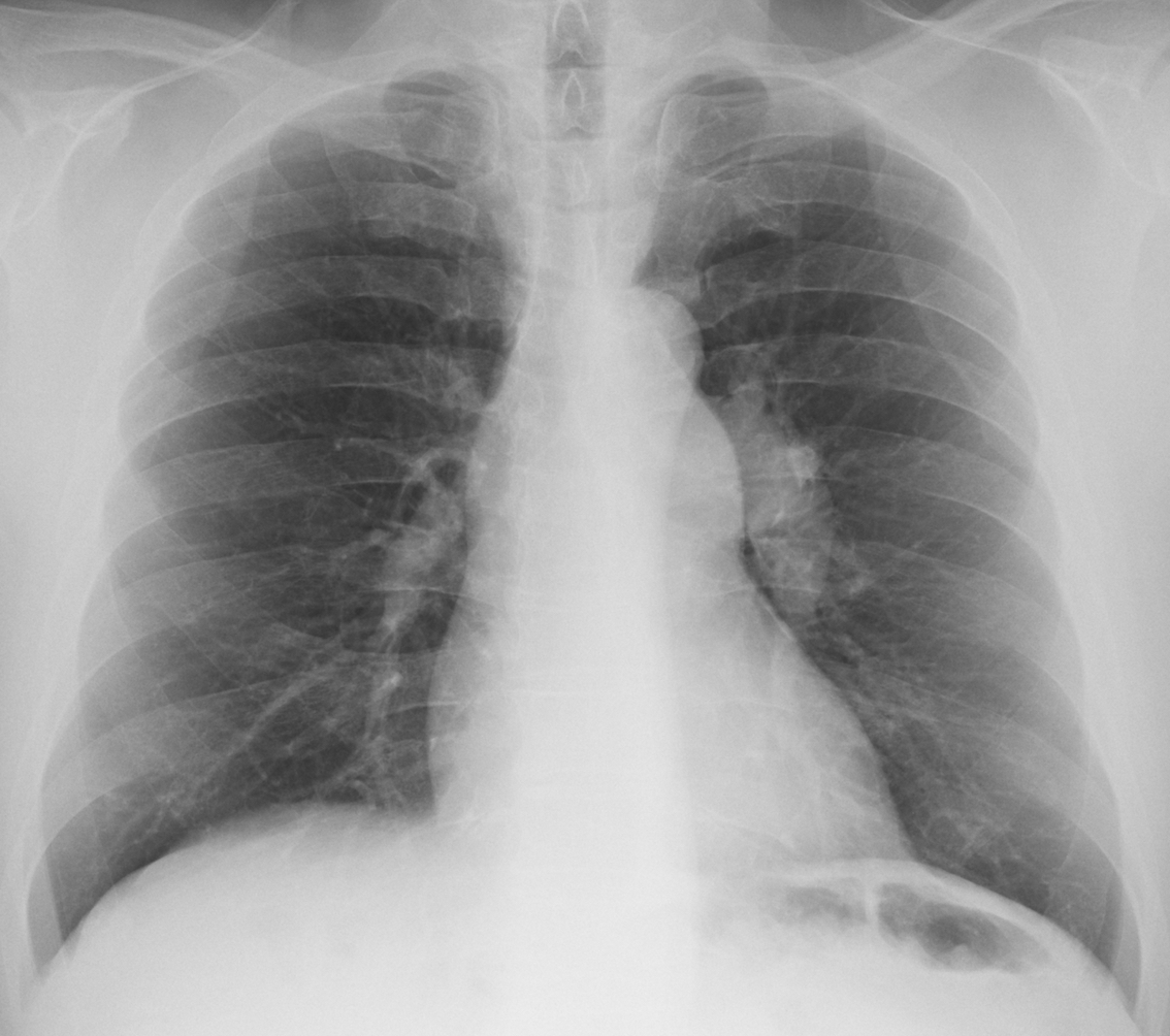
Today we are showing another pre-op case, including a PA chest radiograph of a 62-year-old man with lumbar hernia. Check the image below, give us your thoughts in the comments, and come back on Friday for the answer.
1. TB
2. Carcinoma
3. Pulmonary hypertension
4. None of the above
Findings: the PA radiograph shows a prominent pulmonary artery segment (A-B, arrow) and an enlarged left hilum (A-B, red arrow).
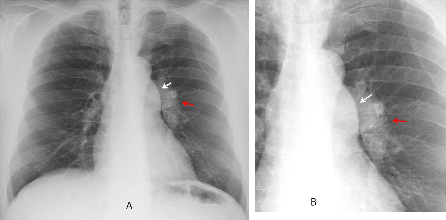
This combination of findings is highly suggestive of dilatation of the main and left pulmonary arteries and should not be confused with enlarged lymph nodes (See Diploma case 52).
Enhanced axial and coronal CT confirms the diagnosis (C-D, arrows).
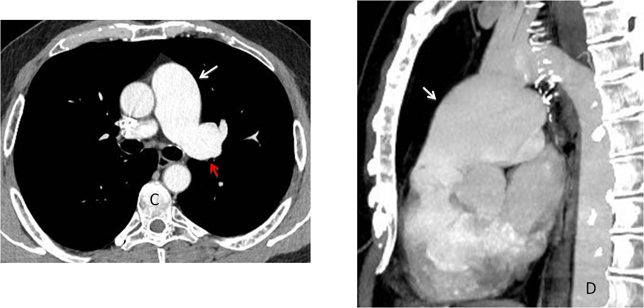
The etiology of these findings is either pulmonary valve stenosis or idiopathic dilatation of the pulmonary artery. In this patient, further work up did not demonstrated valve stenosis.
Final diagnosis: idiopathic dilatation of pulmonary artery
Congratulations to Mauro, who was the first to mention the correct diagnosis.
Teaching point: as I have mentioned before, congenital malformations in adults are not unusual. We should learn to recognise them to avoid confusing them with other pathology.