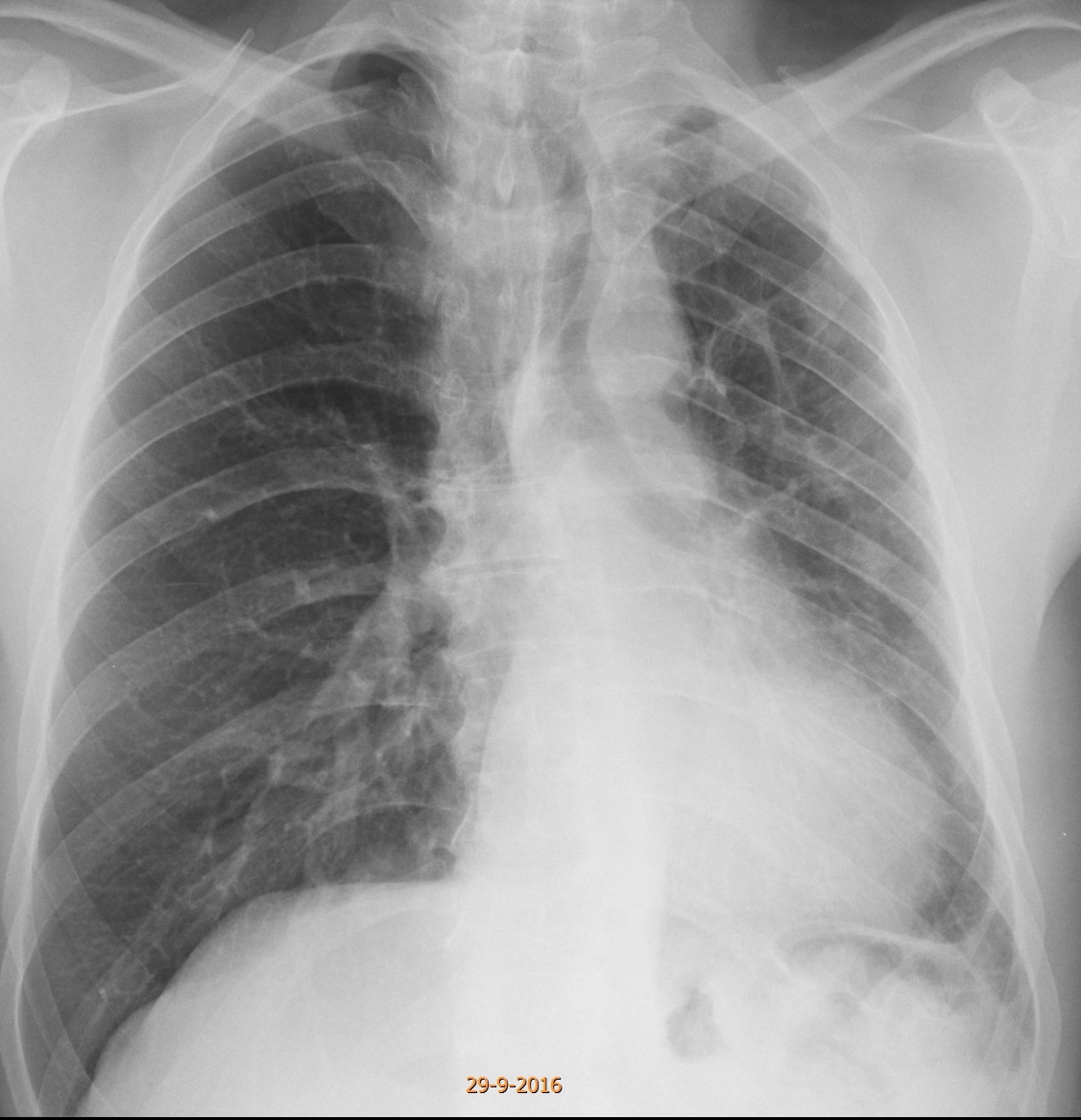
Today we are presenting a recent case. The PA radiograph was made for routine check up of a 49-year-old male.
Check the image below, leave your thoughts in the comments and come back on Friday for the answer.
1. McLeod syndrome
2. Changes post-TB
3. Congenital lung hypoplasia
4. None of the above
Findings: the PA radiograph shows a small left lung, with mediastinal shift (A, arrow). Of the proposed answers, hypoplasia of the left lung is very rare (agenesis occurs more frequently, but it appears as a white hemithorax). McLeod is a good possibility, but an expiration film did not show air trapping. The patient had been diagnosed with old TB in a previous film five years earlier (B), but we questioned him and he denied any history of TB.
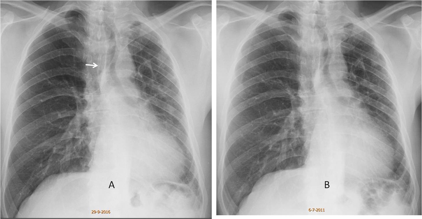
The appearance of the chest and the lack of symptoms suggested to us a congenital vascular hypoplasia, either arterial or venous.
Enhanced CT shows an absent left pulmonary artery (C, arrow), with numerous bronchial collaterals at the left hilum (D, arrow). There is a reticular pattern in the left lung with pleural nodules (E, circle), which represent collateral circulation from pleural vessels.
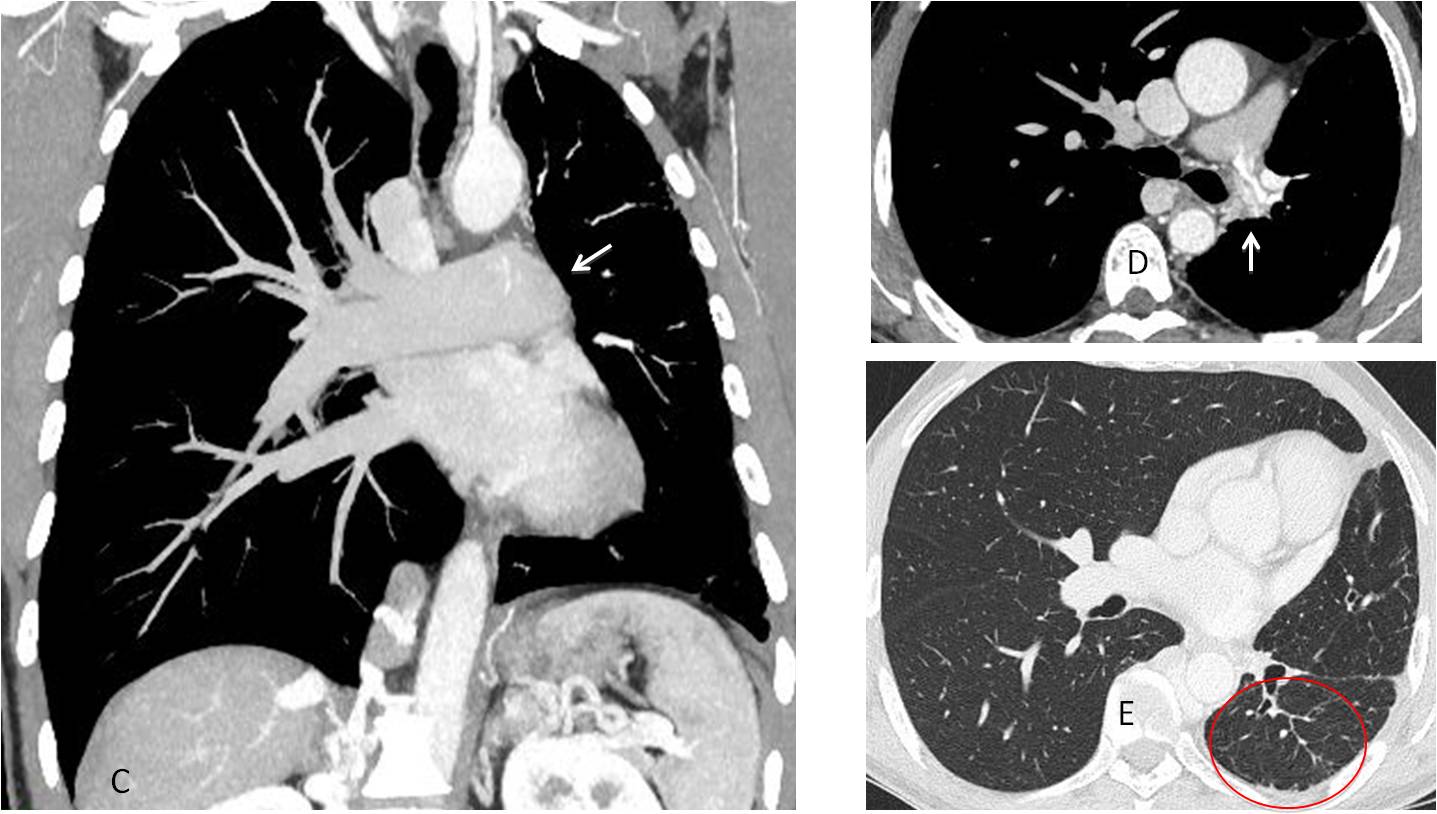
Final diagnosis: congenital absence of left pulmonary artery
Congratulations to Mahmoud and MK, who were the first to suggest this difficult diagnosis
Teaching point: this is another example of a rare congenital condition discovered because intellectual curiosity led us to perform CT and arrive at the correct diagnosis