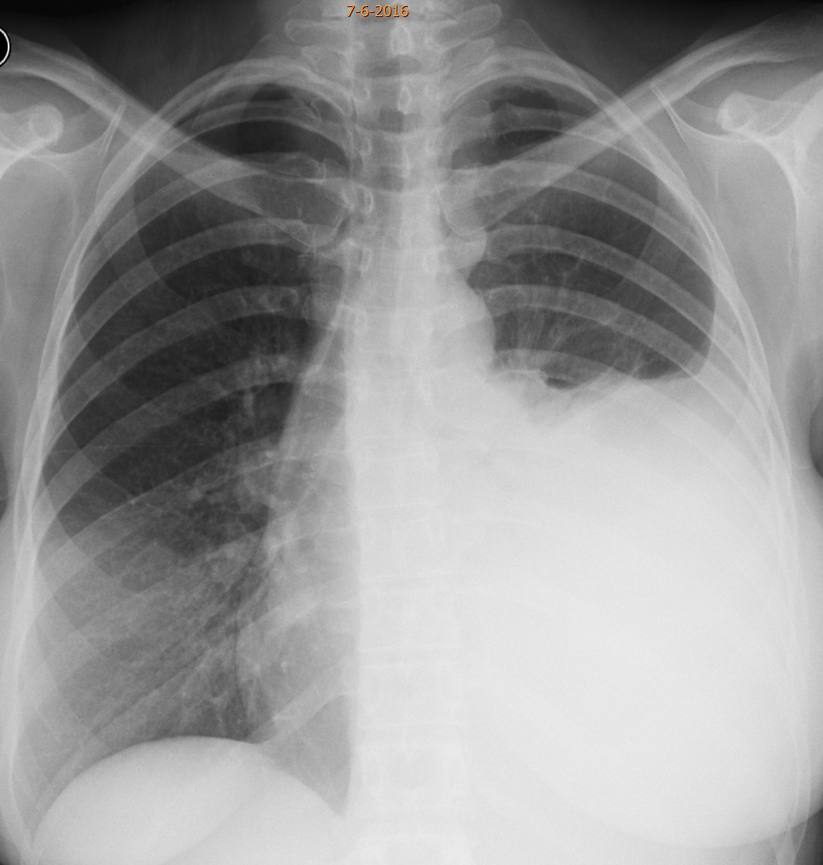
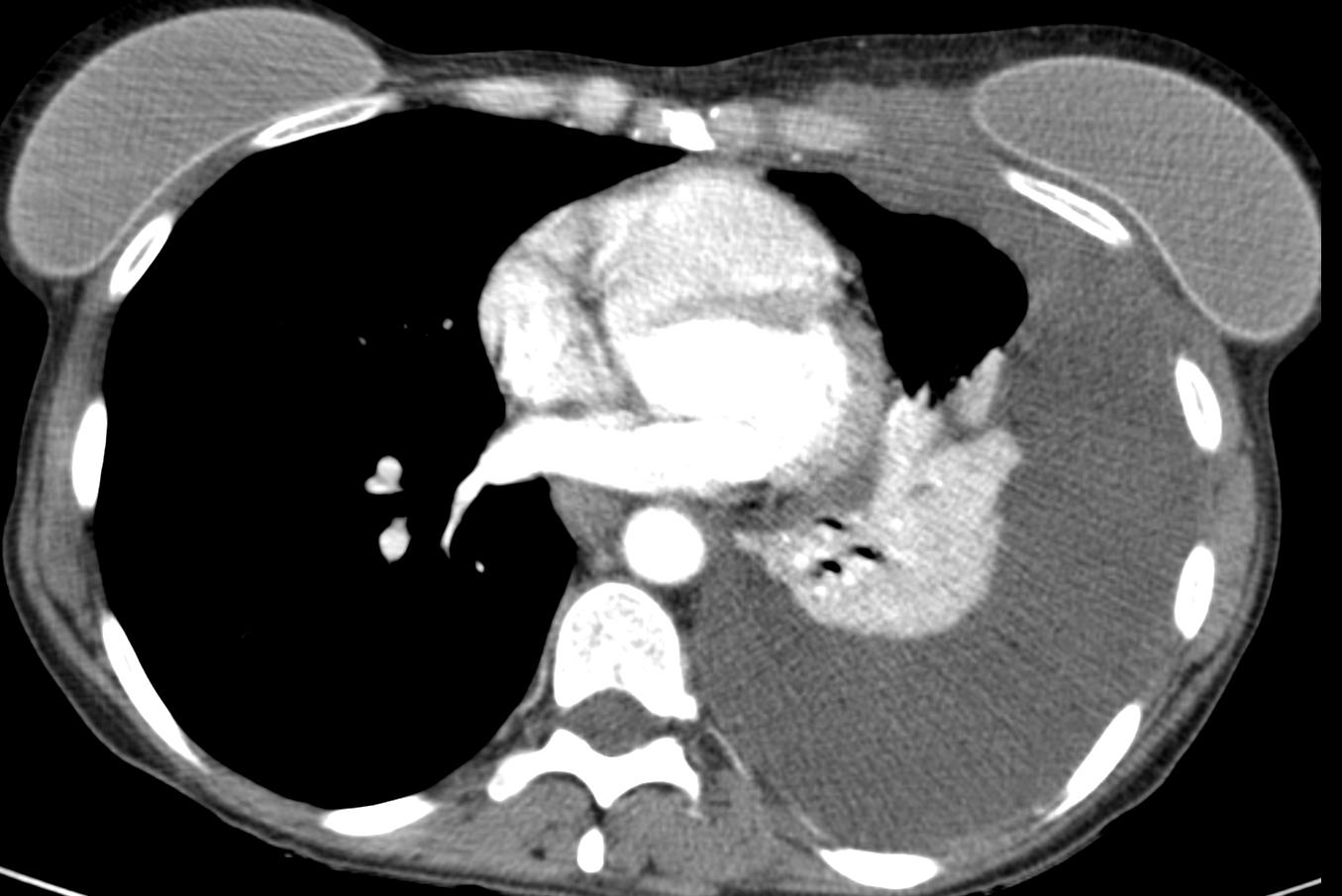
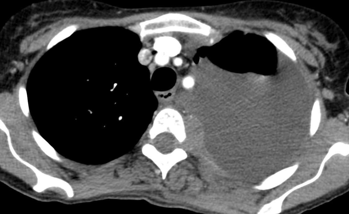
Below are images of a 35-year-old woman with chest pain and progressive dyspnoea for the last three weeks. Leave your thoughts in the comments section and come back on Friday for the answer.
1. Lymphoma
2. Pleural metastases
3. Mesothelioma
4. Any of the above
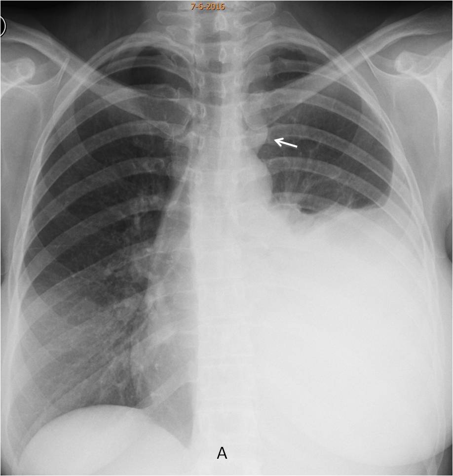
Findings: the PA radiograph shows a large left pleural effusion. A small mass visible in the left upper mediastinum (A, arrow).
Enhanced axial CT confirms the upper mass (B, arrow), which is invading the neural canal. There is a second mass involving the chest wall (C, arrow), adjacent to the left implant, better seen after the fluid was drained (D, arrow).
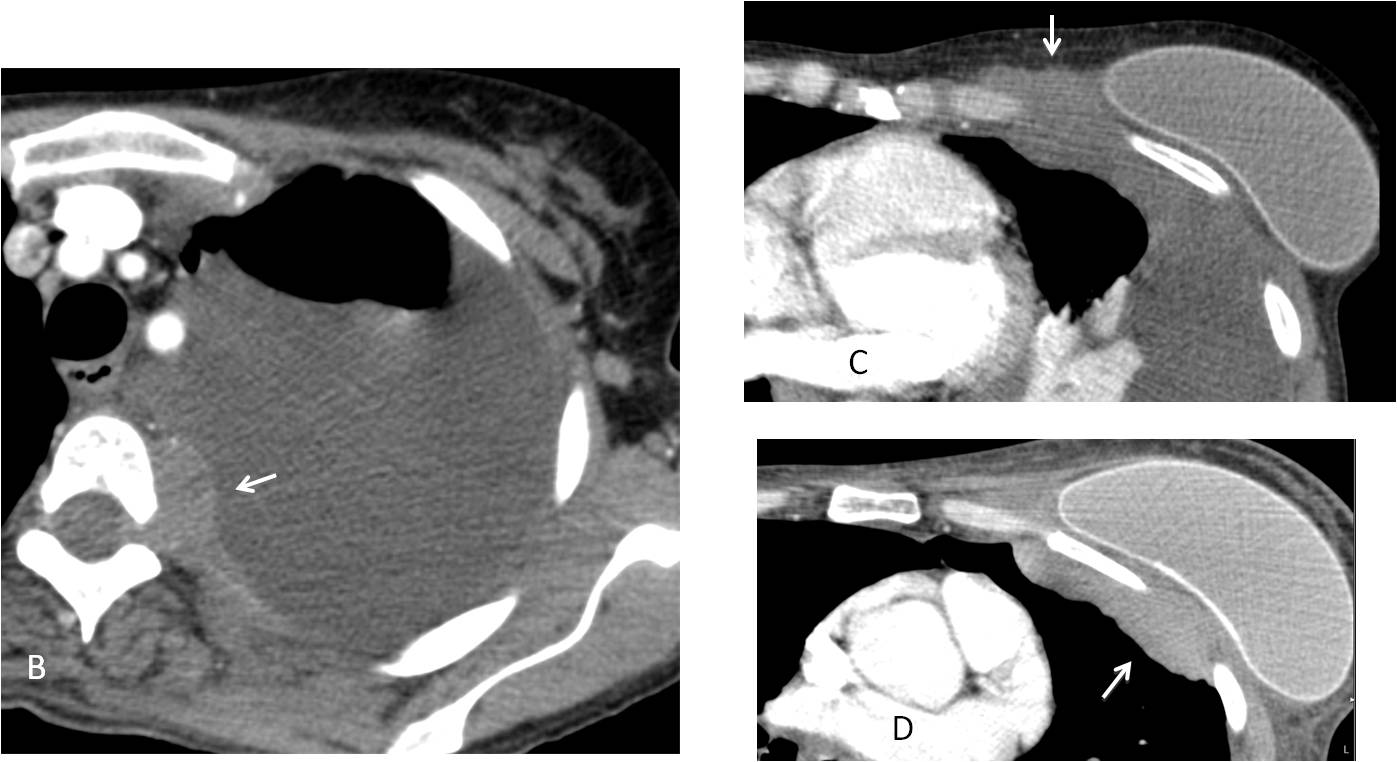
I am showing this case because it is a good example of a little known entity: lymphoma developing in women with breast implants. The relationship is not clear, but all reported cases are anaplastic large cell lymphomas, ALK negative.
Our case had these characteristics. The patient had recurrent seromas in the left breast. Cytological examination of the fluid was negative until the patient developed the actual tumour.
The chest radiograph (E) became normal after treatment.
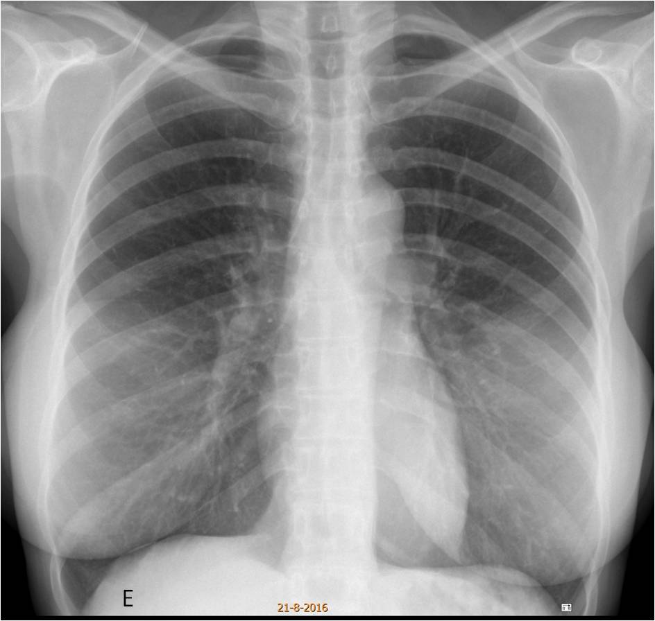
Final diagnosis: anaplastic large cell lymphoma in a patient with breast implants.
Congratulations to Mauro who made a good differential diagnosis, with Mahmoud as a worthy second.
Teaching point: it is wonderful to learn new things! I was shown this case recently by a junior staff and, of course, I had no idea it existed. Whether or not the relationship between implants and lymphoma is real, I am going to look at it from now on. I remember publishing a paper about two patients with implants and recurrent breast seromas and I am planning to review the clinical histories to see what happened.