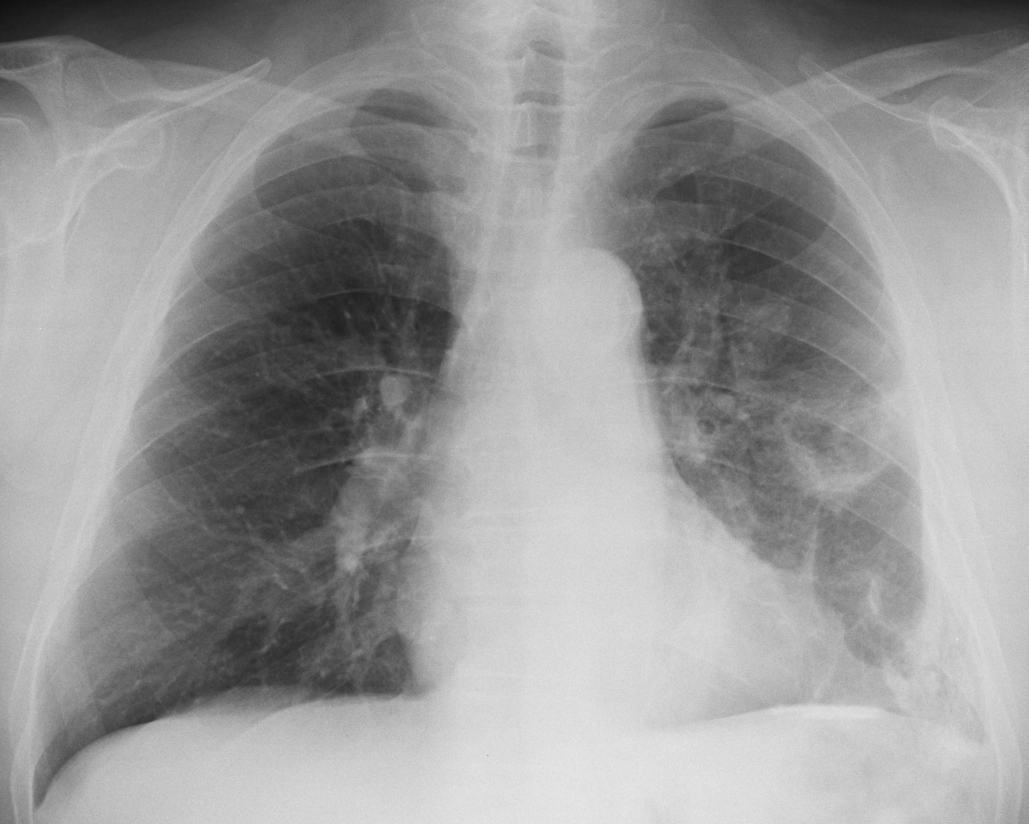
Today we are presenting a pre-op chest radiograph of a 70-year-old man with carcinoma of the bladder.
What do you see?
Check the image below, leave your thoughts in the comments section and come back on Friday for the answer.
The preoperative chest radiograph shows left diaphragmatic and pleural calcifications (A, arrows). There are several opacities in the left hemithorax that may be pulmonary infiltrates (A, red arrows).
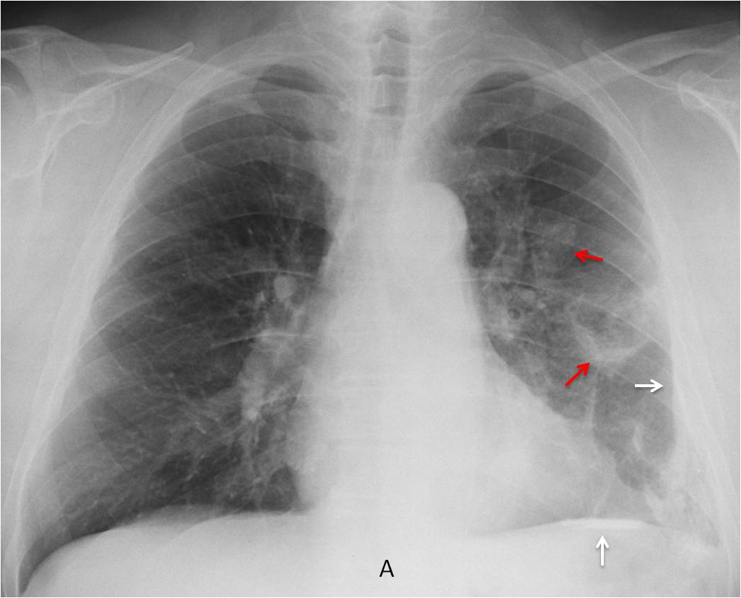
Two considerations should me made: 1) Asbestos exposure is very unlikely because the pleural calcifications are unilateral. 2) The apparent pulmonary infiltrates could be due to pleural calcifications depicted en face.
Coronal CT (B) does not show any lung abnormalities. Axial and sagittal enhanced CT shows extensive pleural calcification (C-D, arrows).
The patient had antecedents of TB in his youth.
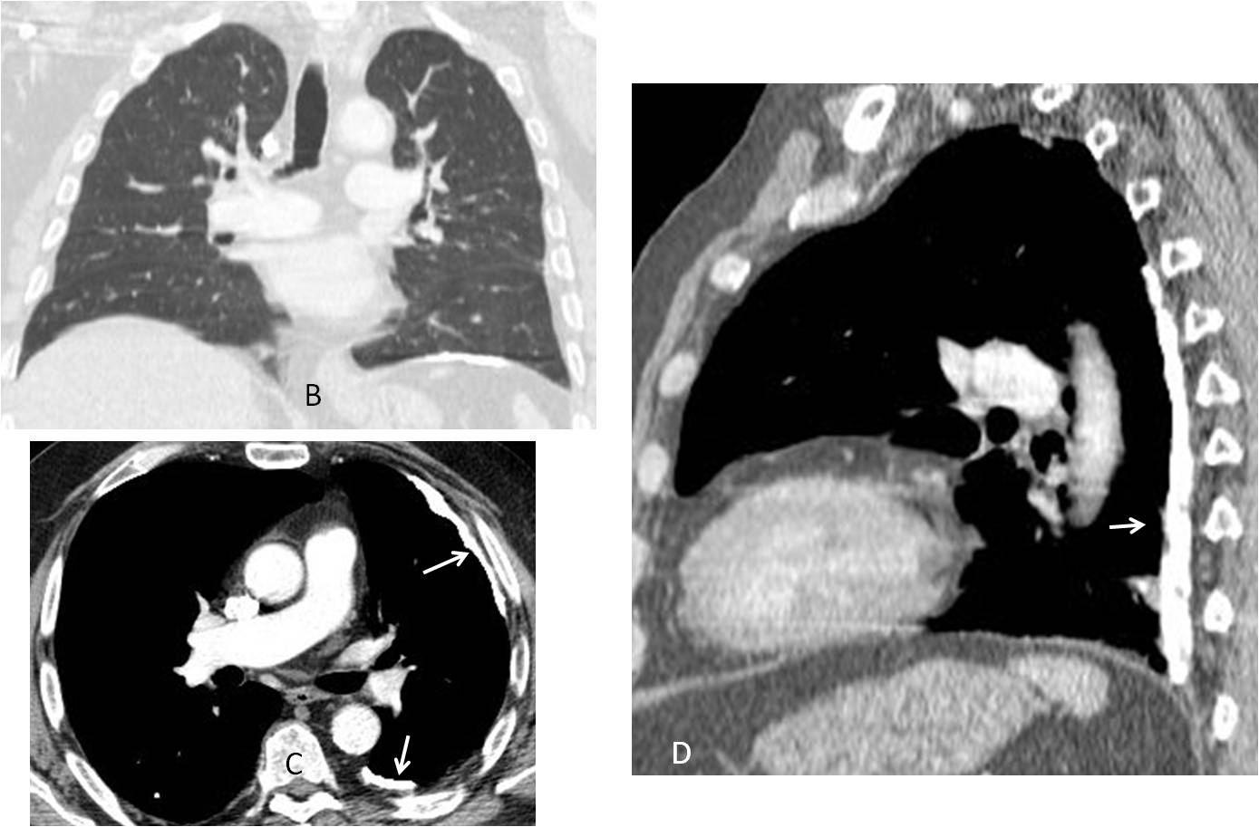
Final diagnosis: Calcified pleura secondary to previous TB
Congratulations to Nacer, who was the first to suggest the diagnosis
Teaching point: Remember that diseases of pleura/chest wall may simulate pulmonary lesions in the chest radiograph