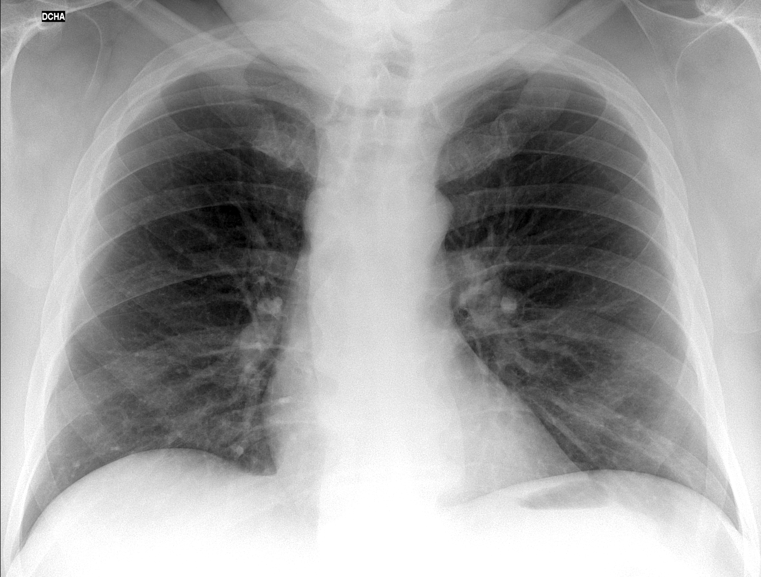
Today we are presenting a pre-op PA chest radiograph of a patient with inguinal hernia. What do you see?
Check the image below, leave your thoughts in the comments section and come back on Friday for the answer.
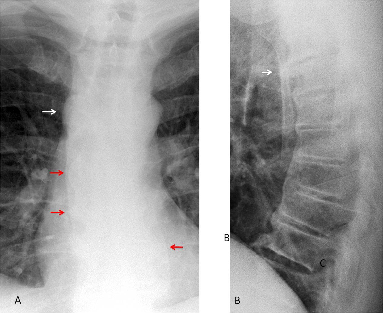
Findings: the PA radiograph shows a rounded shadow in the right upper mediastinum (A, arrow). All mediastinal masses look the same in the plain film; but the clue to the diagnosis lies in the presence of big osteophytes on the right and left sides of the spine (A, red arrows), suggesting that the rounded shadow also represents a big osteophyte. A lateral view taken two years ago shows osteophytes at the same level (B, arrow), accompanied by calcification of the anterior ligament.
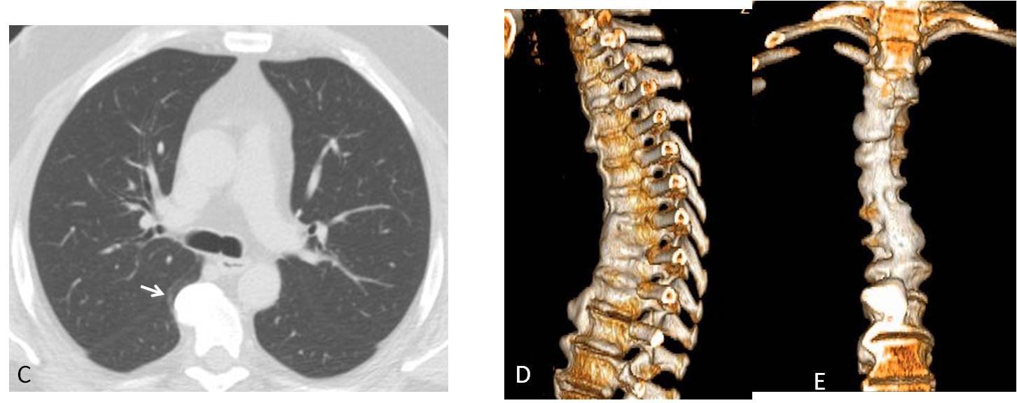
Axial CT shows that the mass corresponds to a large osteophyte (C, arrow).
3-D reconstruction shows extensive calcification that seems to flow along the anterior aspect of the spine (D-E). This appearance suggest a possible DISH
(Forestier disease).
Final diagnosis: osteophyte simulating a mediastinal mass in a patient with probable DISH (unproven).
Congratulations to ZHW, who was the first to suggest the diagnosis.
Teaching point: do not forget that a large osteophyte may simulate a posterior mediastinal mass. And in this patient led to a probable diagnosis of systemic bone disease.