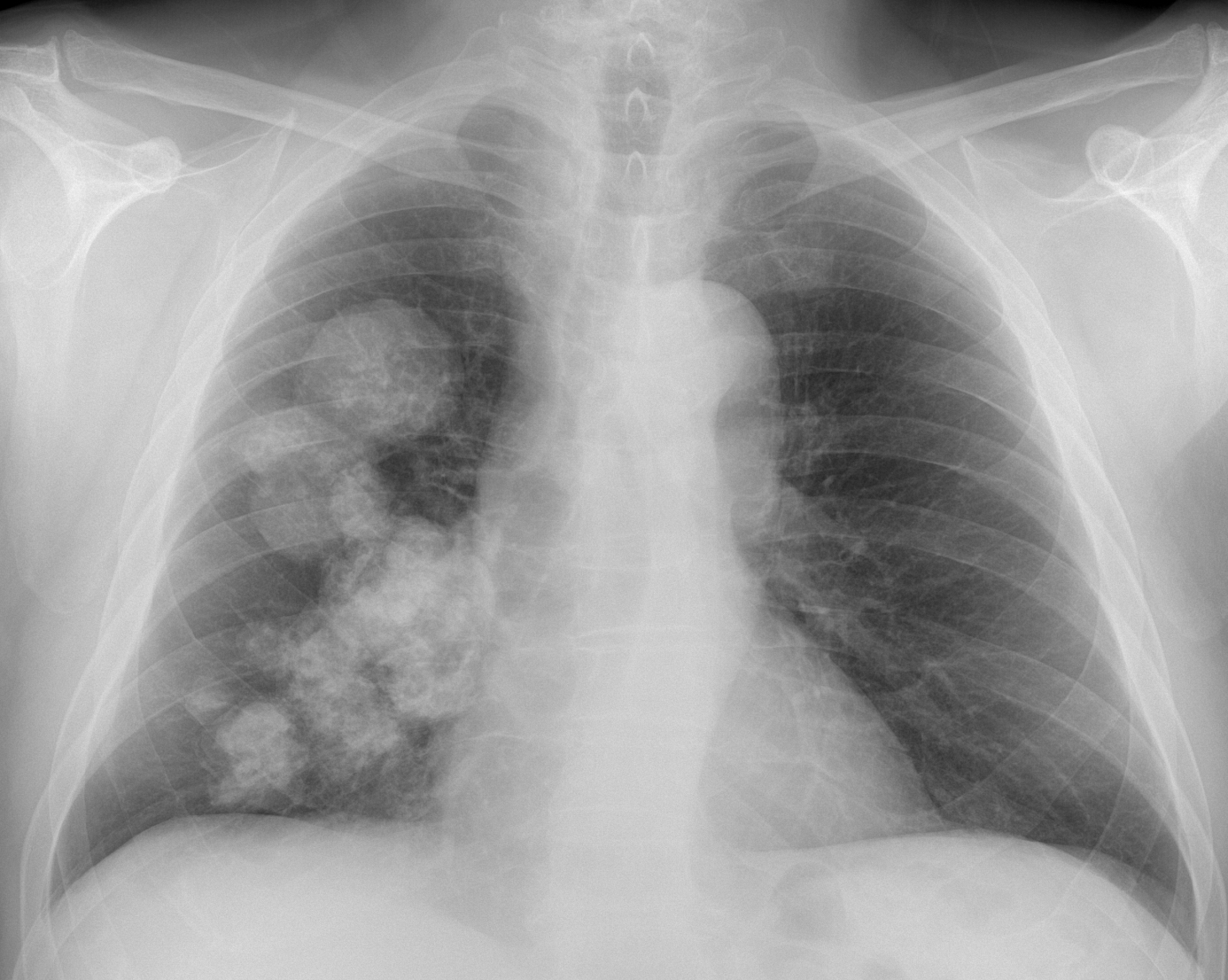
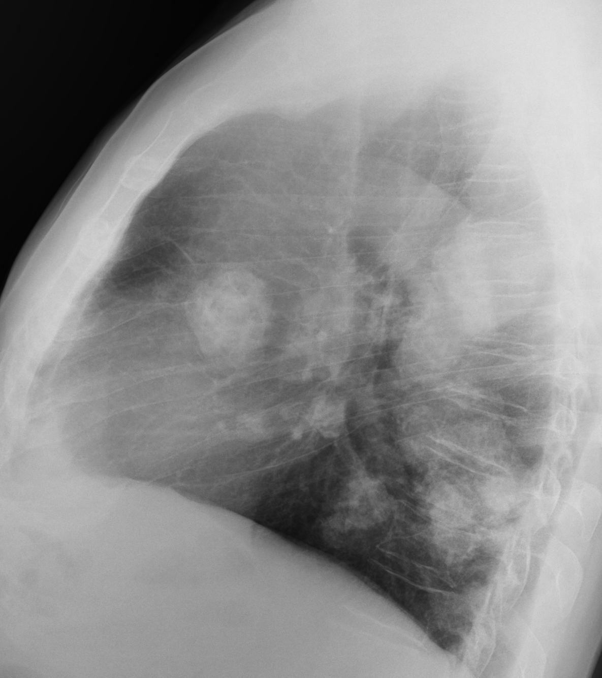
What do you see? Check the images below, leave your thoughts for us in the comments section, and come back on Friday for the answer.
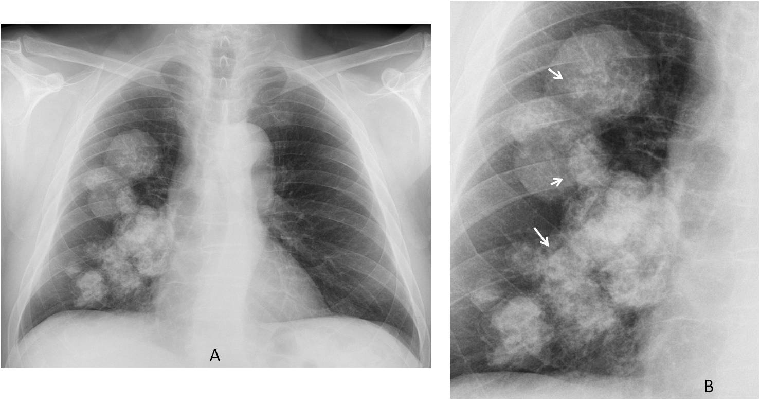
Findings: the chest radiograph shows numerous calcified masses in the right lung (A). The type of calcification is amorphous (B, arrows) and the first impression that came to mind is chondromatous tumours or amyloid. I believe the unilateral location goes against metastases.
I spoke to the patient (highly recommendable technique!) and he said that an oncologist was treating him for an abdominal tumour. I called the oncologist, who confirmed a previous GIST. He told me that the lung lesions had been stable for the last ten years (C, D) and that the clinical diagnosis was Carney’s triad.
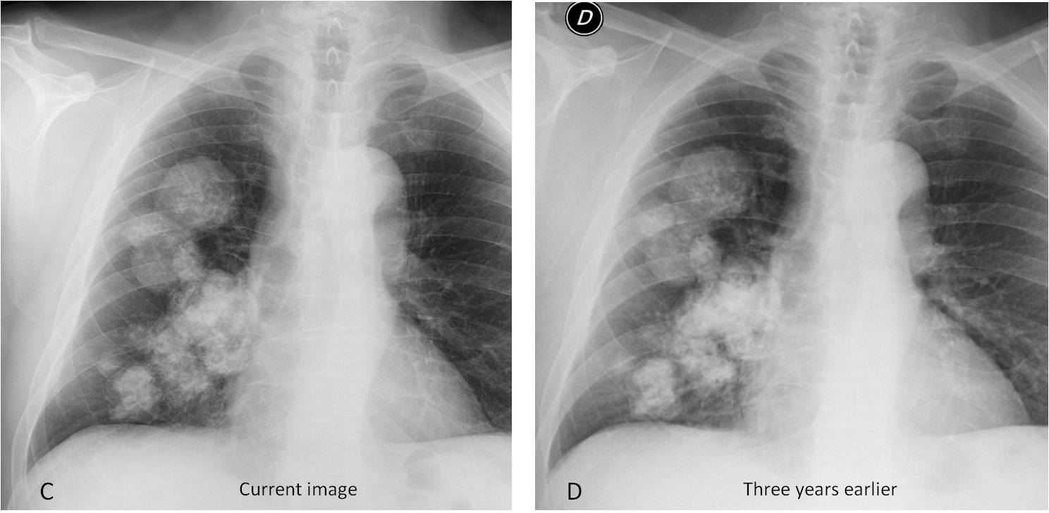
Final diagnosis: Pulmonary chondromas in a patient with Carney’s triad.
Congratulations to Genchi Bari, who was the first to mention multiple chondroid tumours and MK, who jumped to the right diagnosis with a little help.
Teaching point: I got the diagnosis of this unusual case after speaking to the patient. Don’t forget that the patients are an important source of information!
This is the last case of 2016. Dr. Pepe and I are leaving on vacation and will be back on January 9 with new cases.
Happy Holidays to you all!