
Dear Friends,
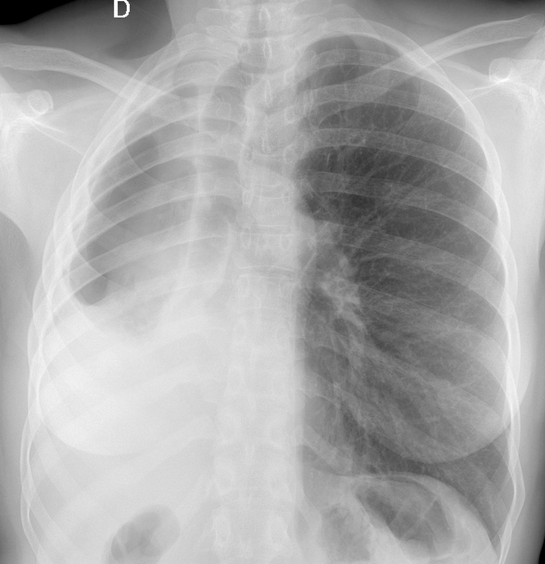
We are starting the new year with a warm-up case. Today we are presenting a PA chest radiograph of a 57-year-old woman with a cough and sputum production.
Check the image below, leave your thoughts in the comments section, and come back on Friday for the answer.
Diagnosis:
1. Changes post TB
2. Congenital right lung hypoplasia
3. Mesothelioma
4. None of the above
Click here for the answer to case #151
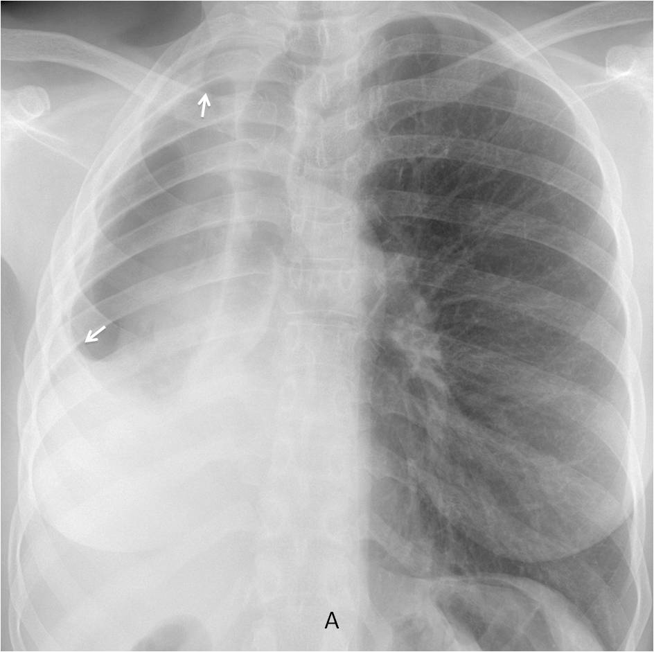
Findings: the PA radiograph shows a small right hemithorax with deviation of mediastinum towards the right. There is marked pleural thickening (A, arrows), and a small right lung. Close inspection shows a lack of vessels in the apparent right lung indicating that the air is in the pleural space. This combination of pleural disease and pleural air is strongly suggestive of a bronchopleural fistula (BPF).
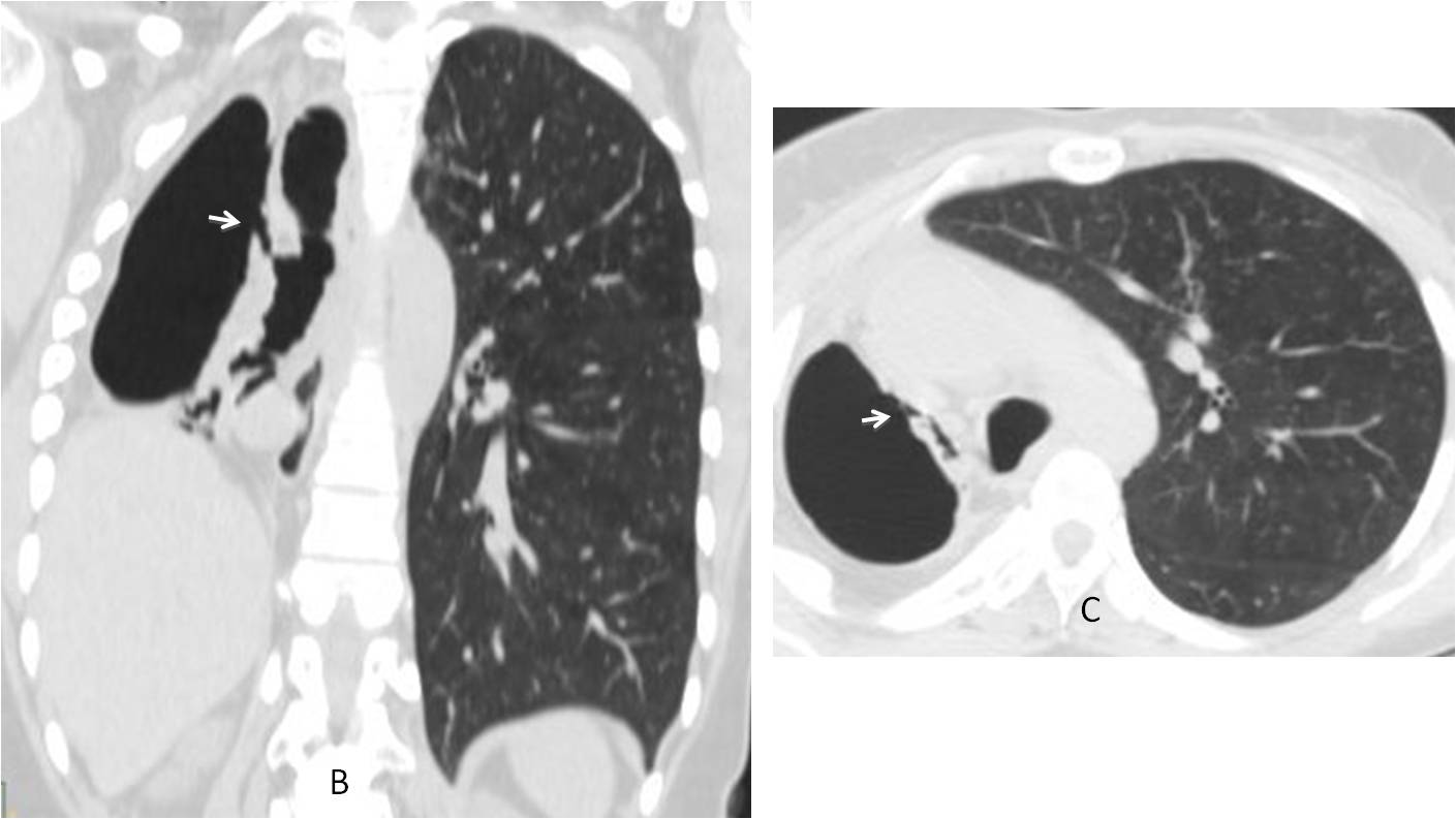
Coronal and axial CT confirm the initial diagnosis, demonstrating the fistulous tract (B-C, arrows). There is marked collapse of the right lung.
The patient was originally from Africa and had had no previous interventional procedures. She did not come back to our institution and was lost to follow-up.
I am showing this case because I thought it interesting to show a rare case of non-surgical BPF.
Final diagnosis: bronchopleural fistula, probably secondary to previous TB
Congratulations to MK who was the first one to suggest the diagnosis
Teaching point: The combination of chronic pleural changes and pleural air with no previous intervention should raise suspicion of BPF.


