Dear Friends,
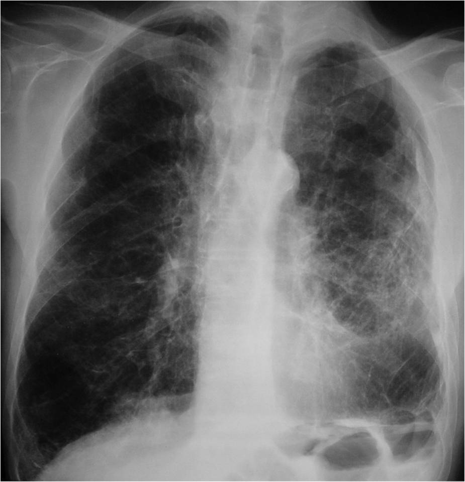
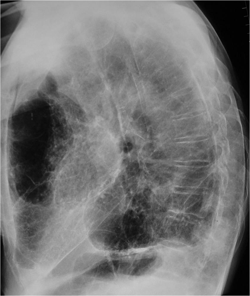
Today we are showing chest radiographs of a 75-year-old man with a cough and haemoptysis.
What do you see? Check the images below, leave your thoughts in the comments section, and come back on Friday for the answer.
Click here for the answer to case #152
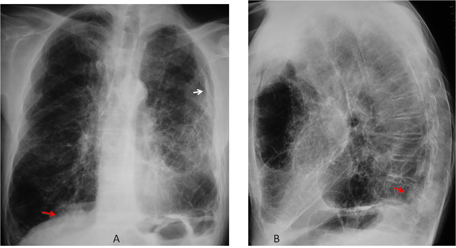
Findings: PA view shows emphysematous lungs with chronic fibrotic changes on the left and pleural calcification (A, arrow). The most important finding is a hidden mass in the RLL, barely visible in the PA radiograph (A, red arrow) and well seen in the lateral view in the posterior costophrenic angle (B, red arrow).
Coronal and sagittal CT confirms the presence of the mass (C-D, red arrows).
Percutaneous biopsy confirmed malignancy.
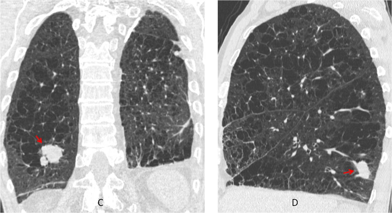
Final diagnosis: partially hidden adenocarcinoma of lung
Congratulations to Mahmoud, who was the first to see the carcinoma
Teaching point: remember the basics. Using checklists helps to find pathology that will be overlooked otherwise.


