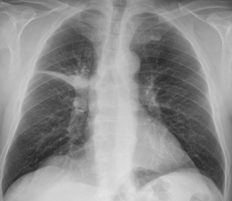
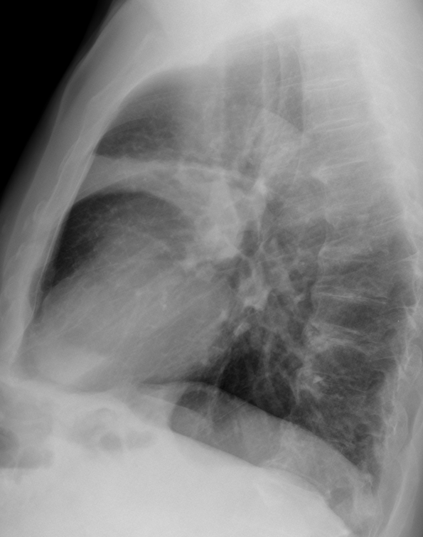
This week we are showing a relaxing case. Images belong to a 64-year-old man with a cough and fever. What do you see?
Check the images below, leave your thoughts in the comments section, and come back on Friday for the answer.
Findings: chest radiographs show a pulmonary infiltrate in the anterior segment of the RUL. There is loss of volume as demonstrated by concavity of the minor fissure (A-B, arrows). For those of you that have diagnosed fluid in the minor fissure, remember that the border is convex instead of concave.
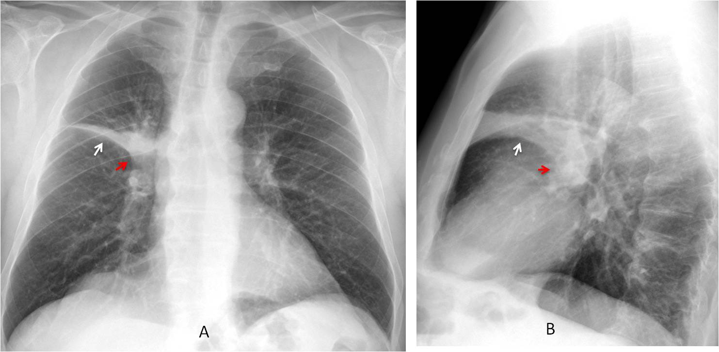
Another significant finding is a mass in the hilar region (A-B, red arrows), creating an evident reverse “S” (Golden sign). This sign is highly suspicious of a central carcinoma.
Enhanced coronal and axial CT demonstrate a mass obstructing the RUL bronchus (C-D, red arrows) and narrowing the main right bronchus (C, arrow). PET-CT shows enhancement of the mass (E, arrow).
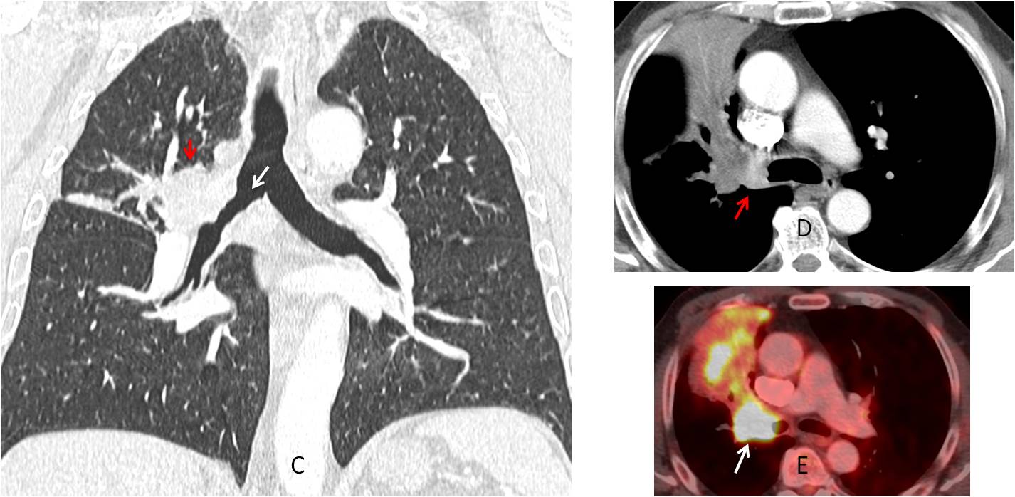
Final diagnosis: carcinoma of RUL bronchus with segmental collapse.
Congratulations to MK, who was the first to suggest the diagnosis
Teaching point: Golden sign is infrequent but, when present, is a good indicator of malignancy and should prompt a CT